Custom Search
Showing posts with label MEDICAL PHOTOS/PICTURES/IMAGES. Show all posts

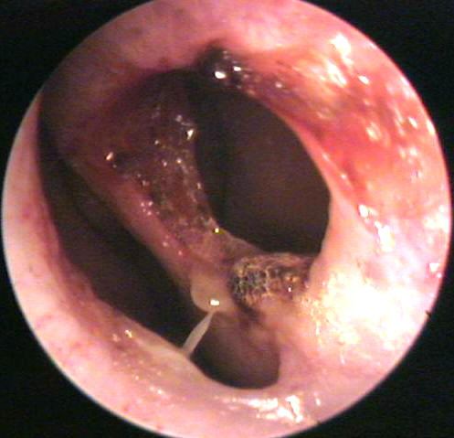
Tongue`s surface in Prolonged antibiotic use

Which of the following conditions is the skin finding shown in the picture associated with?

  • A) Prolonged antibiotic use
  • B) Sjögren's syndrome
  • C) Addison's disease
  • D) Chronic gastroesophageal reflux
  • E) Malignant melanoma


The answer is A. 
(Black tongue) Black hairy tongue results from hyperplasia of the filiform papillae with deposition of keratin on the surface. The condition causes the tongue to have ..............

Types of Hypospadias


Hypospadias is one of the most frequent male congenital malformations and may be part of the testicular dysgenesis syndrome, It occures 1-2 of 100 boys. Hypospadias is a birth defect found in boys in which the urinary tract opening is not at the tip of the penis. Bending of the penis on erection may be associated and is as chordee.


This picture shows Proximal shaft hypospadias . Note the deficient ventral foreskin, blind urethral pit at the glanular level, and lighter pigmented urethral plate extending to the true meatus at the proximal shaft level.

The location of the urethral meatus was described if visible, and hypospadias was graded as glandular, coronal, penile, penoscrotal, scrotal, or perineal according to the anatomical position


Types of hypospadias classified by the anatomical position of the urethral meatus. 1, Glandular; 2, coronal; 3, penile; 4, penoscrotal; 5, scrotal; 6, perineal.

On examination :A dorsal hood of foreskin and glanular groove are evident, but, upon closer inspection, the prepuce is incomplete ventrally and the urethral meatus is noted in a proximally ectopic position. Rarely, the foreskin may be complete, and the hypospadias is revealed at the time of circumcision. If hypospadias is encountered during neonatal circumcision, after the dorsal slit has been performed, the procedure should be halted, and the patient should be referred for urologic evaluation.
 The most simple classification of Hypospadias is mild, moderate and severe, which can also be called first, second and third degrees of severity. This classification system is based on the location of the external opening for urine and semen (the urethral meatus). In mild or first degree hypospadias, the opening is on the underside of the head of the penis or where the head and the shaft meet. This accounts for about 80% of the cases of hypospadias.

Moderate or second degree hypospadias (15%) occurs when the hole is actually on the shaft of the penis, somewhere between the junction of head and shaft, and the lower part of the shaft. Severe or third degree hypospadias occurs when the hole is located on the lower part of the shaft just in front of the testicles or is located behind the testicles, between them and the anus.

Photos of Melanosis coli "Pseudomelanosis coli"

Melanosis coli, also pseudomelanosis coli, is a disorder of pigmentation of the wall of the colon, often identified at the time of colonoscopy.
The most common cause of melanosis coli is the extended use of laxatives, this darkening of the colonic mucosa caused by the accumulation of lipofuscin particles within the macrophages of the lamina propria of the colonic mucosa as a result of long term exposure to anthraquinone-containing laxatives as Senna and other plant glycosides. However, other causes are identified, including an increase in colonic epithelial apoptosis.

This is the photograph of the colon, and there is an extremely dark appearance to the wall of the colon. This is seen in patients who have taken laxatives over many years and the pigment from the laxative gets deposited in the wall of the bowel giving an extremely dark appearance to it. This is a benign condition, not cancerous, and does not become cancerous, but often it is quite obvious.

- Melanosis coli is a misnomer, as the pigmentation is due to lipofuscin-laden macrophages - not melanin pigment. Pseudomelanosis coli is a more appropriate descriptor, but not in common usage.

The differential diagnosis of brown pigmentation of the colon is:

* Pseudomelanosis coli.
* Hemosiderin-laden macrophages (old haemorrhage).
* Melanin (rare).

Description of Hallux Malleus "hammer toe"

-Hallux for the big toe
-Malleus for a hammer

Hallux malleus is a deformity of the great toe. This deformity can be very stiff or flexible. The joint in the great toe becomes contracted in a flexed or downward position.
This deformity usually occurs due to an imbalance of the tendons that insert on the top and the bottom of the great toe. When the tendon on the bottom of the toe (the tendon that causes the toe to flex down) over powers the tendon that causes the toe to bend up, this deformity occurs.
Usually patients will develop a callus and even an ulcer on the tip of the great toe. This deformity is often seen in conjunction with hammer toes. High arched feet are typically the most affected by this deformity.

This deformity is treat initially with padding techniques to prevent sores from developing at the tip of the toe. Custom, soft, accommodative orthoses are sometimes prescribed for the patient to provide cushioning and also to prevent worsening of the deformity.

If conservative treatment fails, surgery is indicated. Surgery usually consists of performing a bone fusion of the 2 bones in the great toe. This can be done with screws, staples, or wire fixation.

Photo illustration of Actinic Keratosis

Actinic keratoses generally appear as rough, red/brown, scaly macules or papules on the skin. They start to appear usually about age 30 or older.


Actinic keratoses (AKs) are A premalignant neoplasm of the epidermis caused by excessive exposure to sunlight and manifesting as an ill-marginated, erythematous, scaling, rough papule or patch that form on sun exposed areas of the skin, including the scalp, face, forearms, and back of the hands.

Actinic keratoses are usually scaly, feel like sandpaper to the touch, and range in color from skin-toned to reddish-brown " AK are more easily felt than seen, as their overlying scale is thick and firmly adherent".  Actinic keratoses may be as small as the head of a pin or as large as a quarter (or bigger, if left untreated).
Actinic keratoses on the scalp

Actinic keratoses are most common in individuals over 40 years old with fair skin and years of excessive sun exposure. However, even younger people (including those with dark skin) can develop actinic keratoses if they live in very sunny climates.

Lesions are occasionally tender to palpation. Fair-skinned persons, who burn easily and tan poorly, are most commonly affected. A small percentage of AK on non-mucosal skin can progress to skin cancer squamous cell carcinoma. Ultraviolet light exposure induces formation of the lesions.

How to distinguish Athetosis from Chorea ?

Sometimes is difficult to distinguish athetosis from chorea (hence the term choreoathetosis). Typically, however, athetoid movements are slower than choreiform movements.

Athetosis is an extrapyramidal sign characterized by slow, continuous, twisting, involuntary movements. Typically, these movements involve the face, neck, and distal extremities, such as the forearm, wrist and hand.
Facial grimaces, jaw and tongue movements, and occasional phonation are associated with neck movements. Athetosis worsens during stress and voluntary activity, may subside during relaxation, and disappears during sleep. It is commonly a lifelong affliction.
A: With athetosis, movements are typically slow, twisting, and writhing. They're associated with spasticity and most commonly involve the face, neck, and distal extremities. 
B: With chorea, movements are brief, rapid, jerky, and unpredictable. They can occur at rest or during normal movement. Typically, they involve the hands, lower arm, face, and head.

Positions of fingers in movements of athetosis.
Athetosis usually begins during childhood, resulting from hypoxia at birth, kernicterus, or a genetic disorder. In adults, athetosis usually results from a vascular or neoplastic lesion, a degenerative disease, drug toxicity, or hypoxia.

Photo description of Pterygium of the eye

Pterygium is a raised, wedge-shaped, non-cancerous growth of the conjunctiva onto the cornea, characterized by elastotic degeneration of and fibrovascular proliferation
Pterygium has a characteristic shape as it is triangular, with the base of the triangle located in the conjunctiva and the apex of the triangle encroaching onto the cornea.
With corneal involvement, even if arrested surgically, a pterygium can affect vision by warping the surface of the cornea and inducing astigmatism, and/or by actually growing over in front of the pupil and obstructing the entering light.
 

Triangular, thin, transparent conjunctival fold in the palpebral fissure. Its head is yellow, avascular and pints toward the corneal center. The body of the pterygium extends to the semilunar fold(this image for a 32-year-old man).

Treatment :
Pterygia seem to occur more frequently in people who spent much time outside, and is especially common in the southern lattitudes "Some consider pterygium as an occupational disease" So protecting the eyes from sun, dust and wind and use of lubricating drops is the standard treatment.
If the pterygium is still troublesome it can be removed surgically under local anaesthetic ,with high rate of recurrence. Once the pterygium is removed, the bare sclera is covered with conjunctiva

Photos and information about Blue Nevus

- Blue Nevus is a group of melanocytic lesions which all appear blue in colour due to the optical effects of light reflecting off melanin deep in the dermis.

- The blue nevus is a benign, usually solitary lesion, represents a localized proliferation of dermal melanocytes. It presents as a dark blue to black, moderately firm, rounded, sharply defined nodular tumour composed of spindle-shaped melanocytes with slender cytoplasmic processes, occurring often in association with melanin-laden macrophages in a sclerotic dermis.
- A blue naevus (nevus) is a rather unusual but non-cancerous mole.

- The blue nevus is also called the blue skin mole, or Jadassohn-Tièche nevus with 2 clinically recognized variants : the common blue nevus and the cellular blue nevus.
- Cellular blue nevus is larger, especially on buttocks and can degenerate into malignant melanoma.

- Clinically The blue nevus appears as a dark-blue or blue-black smooth nevus formed by melanin-heavily pigmented spindle cells in the middle and lower two-thirds of the dermis. Also, the blue nevus appears as a slate blue or bluish black, sharply circumscribed, flat or slightly elevated nodule, occurring on any area of the body.
- A biopsy should be performed on any changing pigmented lesion. For a solitary lesion, simple excision is usually curative. Rare cases of persistent blue nevi, manifesting as satellite lesions around the original excision site, have been reported. These must be distinguished from malignant blue nevus, and reexcision is recommended.

Common blue nevus

Diastasis Recti in newborns


A vertical bulge down the midline of the abdomen can be seen in many newborns when intra-abdominal pressure increases. In this photo, a shadow lateral to the bulge can be seen going up the left side of the abdomen, starting near the umbilical clamp. Diastasis recti is caused by a relative weakness of linea alba "the fascia between the two rectus abdominus muscles".
It is essentially a cosmetic condition, with no associated morbidity or mortality .As it is not a herniation and is not pathologic, No treatment is needed ..... with time, this will disappear.
Another newborn has more pronounced diastasis recti. Spontaneous resolution is still expected. 

Atypical "Rodent Ulcer" Basal Cell Carcinoma

These lesions are moist ulcers which may not have the characteristic rolled, translucent border of a nodular Rodent Ulcer or Basal Cell Carcinoma BCC. Often, they are much larger than the pre-auricular lesion seen here, and represent neglected lesions.

The typical basal cell carcinoma appears as a small, pearly, dome-shaped nodule with small visible blood vessels (telangiectasias).

Photo illustrations of Bullous Myringitis

What is Bullous Myringitis ?
Bullous myringitis is painful inflammation blisters of the tympanic membrane and surrounding deep canal skin with the formation of serum or blood filled bullae. Petechial subcutaneous hemorrhages around the base of the bulla are characteristic.

Bullous Myringitis was thought that Mycoplasma pnumoniae was the most common pathogen but, recent studies have demonstrated that the pathogens are similar to otitis media. Bacteria are responsible for most cases of bullous myringitis. Streptococcus pneumoniae is the most common, followed by Haemophilus influenzae and Moraxella catarrhalis.

Examination shows a vesicular or bullous eruption over the tympanic membrane and adjacent bony canal wall. In the early stages, the vesicles are erythematous and surrounded by injected epithelium. In the later stages, they are larger and filled with fluid which may be clear and serous or blood filled. In this photograph, the bulla has now been incised with a myringotomy knife and the fluid drained. Notice the petechial hemorrhage around the base of the bulla and extending into the attic area.
Treatment of bullous myringitis requires strong analgesics because of the associated pain. Rupturing the bullae for relief of pain is controversial. Topical antibiotic eardrops are probably useful in preventing the development of a secondary bacterial otitis externa. The use of erythromycin has been advocated by those who believe that Mycoplasma is the prime etiologic agent. The tympanic membrane in this case is diffusely inflamed, with the presence of petechial hemorrhage over the attic and an associated hemotympanum


A large bulla filled with serous fluid has developed on the superficial surface of the tympanic membrane in the region of the umbo

Illustrated photos of Milia


Definition of Milia :
Milia form as multiple, firm, white lesions that range from 1 to 4 mm in diameter. They are very common, benign, keratin-filled cysts.

Milia usually appear on the face and most commonly affect the eyelids, nose, and malar region, sometimes on the genitalia; It is formed at base of hair follicle or sweat gland.
Milia are commonly associated with newborn babies 40% of newborns, but can appear on people of all ages.


Other names: milk spot or an oil seed.Milium is the single of milia.

Types of Milia :
1- Primary or spontaneously.
2- secondary caused by trauma, radiotherapy, skin infection, or bullous diseases.

Treatment:
In children milia often disappear within two to four weeks.Options include simple excision, electrodessication of the surface, or puncture and expression of the contents

Mottled skin or Cutis Marmorata in neonates

Cutis marmorata or "marbled skin" refers to mottled skin, typically in newborns in the first few months of life.
This reticulated pattern of constricted capillaries and venules is often called "mottling" due to vasomotor instability in immature infants. It generally resolves with increasing age and for most infants is of no significance.

Explanation of cutis marmorata :
This phenomenon is caused by instability or immaturity of the nerve supply to the superficial capillary blood vessels in the skin. This causes the blood vessels in some regions of the skin to dilate, producing a red color of the skin, while other regions are contracting, producing pale skin.

In spite of it`s insignificance in most babies ,it may reflect underlying poor perfusion and infants who develop mottling and are unwell need to be clinically evaluated for sepsis and other illnesses.

Cutis Marmorata Telangiectatica Congenita :

Reticular skin lesions on the right arm of a 7-y girl

It is localised , marked and more pronounced form of cutis marmorata which may be associated with other defects. It is uncommon and sometimes called (congenital generalized phlebectasia)
cutis marmorata telangiectatica congenita is often reported in association with a variety of other congenital anomalies, the most common anomaly associated is Body asymmetry.

A case of Chromoblastomycosis

A 55-year-old man presented with vegetating lesions on the right foot that had been slowly enlarging during the past several years ;The photo:
  On physical examination, several nodular and verrucous lesions were seen in the distal region of the foot. The patient lived in a rural area and had walked barefoot for most of his life. Analysis of a skin-biopsy specimen revealed clusters of small, round, thick-walled, brown sclerotic bodies in the stratum corneum (muriform cells), which are diagnostic for chromoblastomycosis.
Chromoblastomycosis is a chronic, soft-tissue fungal infection commonly caused by Fonsecaea pedrosoi, Phialophora verrucosa, Cladosporium carrionii, or F. compacta. The infection occurs in tropical or subtropical climates and often in rural areas. The fungi are usually introduced to the skin through cutaneous injury from thorns, splinters, or other plant debris. The patient was treated with multiple surgical excisions and itraconazole for 24 months with a complete resolution of symptoms.

Skin manifestations of Pellagra

Pellagra is a a vitamin deficiency disease caused by chronic lack of nicotinic acid (niacin, vitamin B3) or its precursor, tryptophan.Niacin is required for most cellular processes.

CAUSE of Pellagra:
Inadequate niacin and/or tryptophan in the diet mainly seen in developing countries or poverty stricken areas "called Primary pellagra".But usually there is something prevents Niacin absorption and processing and causes secondary pellagra such as:
• Alcoholism, malabsorption, anorexia nervosa, Prolonged diarrhoea, Liver cirrhosis, Drugs e.g. isoniazid, azathioprine

• Carcinoid tumors (excessive use of tryptophan, which produces serotonin)

Symptoms and signs of Pellagra is classically described by "the four D's": diarrhea, dermatitis, dementia and If left untreated, death.

Skin manifestations and clinical finding of Pellagra:
-At first there is reddened skin with superficial scaling in areas exposed to sunlight, heat & friction. This may resemble severe sunburn then gradually subsides leaving a dusky brown-red colouration .
-The rash is usually symmetrical with a clear edge between affected and unaffected skin and sometimes may be itching or burning sensation
-Other features sometimes present include cheilosis, glossitis, angular stomatitis, and oral or perianal sores.

Legs and feet of a patient with niacin deficiency. A, Before therapy. B, After therapy.
(From Swartz MH: Textbook of Physical Diagnosis, 5th ed. Philadelphia, WB Saunders, 2006.)

TREATMENT of Pellagra:
• Nutritional supplementation with niacin will improve the appearance of skin lesions

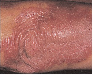
Psoriasis photo affects the elbows

A 45-year-old woman presents with a localized area of erythematous scaly patches that comes and goes and typically affects the elbows. The likely diagnosis is.........
  • A) pityriasis rosacea
  • B) mycosis fungoides
  • C) tinea corpora
  • D) nummular eczema
  • E) psoriasis

The answer is E. (Psoriasis) 
Psoriasis usually manifests itself as erythematous scaly patches that affect the knees or elbows. More severe cases can involve multiple areas over the entire body. Extensor surfaces are predominantly affected. Nail pitting may be present. The condition appears to be hereditary. Diagnosis is usually based on clinical findings. Skin biopsy may be helpful for definitive diagnosis. Treatment consists of topical steroids, intralesional steroids, tar preparations, anthralin, tazarotene, and calcipotriene.

ALCOHOL as Teratogen :Fetal Alcohol Syndrome

In many countries alcohol is the Most common teratogen.And the severity of spectrum of effects in the neonate correlates with the amount of alcohol consumed , ranging from mild reduction in cerebral function to classic fetal alcohol syndrome .

Facial features of Fetal Alcohol Syndrome (FAS)
* Microcephaly - leads to small head circumference
* Palpebral fissure - short opening of eye
* Epicanthal folds - fold of skin at inside of corner of eye
* Midface - flat
* Nasal Bridge - low
* Philtrum - Indistinct, vertical grooves between nose and mouth
* Upper Lip - thin
* Micrognathia - small jaw
* Ears - curve at top part of outer ear is underdeveloped and folded over parallel to curve beneath. Gives the appearance of a "railroad track"

It also associated with :
#Mild to severe mental retardation resulting in learning difficulties.
#Low birth weight and height that persists through early childhood.
#Abnormalities of the heart and other organs.

Fetal Alcohol Syndrome. Notice the depressed nasal bridge, flat philtrum, long upper lip, and thin vermillion border.

Perforation of Nasal Septum


Etiology of nasal septal perforations can be classified into these 4 main categories: traumatic, iatrogenic, inflammatory/malignant, and cocaine use.

Most traumatic or iatrogenic perforations result from mucosal lacerations on corresponding sides of the septum with exposure of the underlying cartilage or from a fracture of the cartilaginous septum. Perforation occurs because the cartilage relies upon the overlying mucoperichondrium for its blood supply and nutrients.
Iatrogenic causes include nasal surgical procedures (prior septal surgery is the most common cause of septal perforations) and nasal intubation or nasogastric tube placement.

Septal hematoma, if not identified and treated early, also may result in perforation.
Infectious and inflammatory etiologies, including tuberculosis, syphilis, Wegener granulomatosis, and sarcoidosis, always should be considered in the differential diagnosis.

Treatment involves the use of nasal wetting agents, placement of a plastic button over the perforation, and surgical repair. Repair with surgery is very difficult and failure to close the perforation is common.

Barrett's esophagus

The Key word :
BARRett's = Becomes Adenocarcinoma, Results from Reflux.

Barrett's esophagus is a Glandular metaplasia ,or replacement of non keratinized (stratified) squamous epithelium with intestinal (columnar) epithelium in the distal esophagus as a serious uncommon complication of chronic acid reflux or GERD (gastroesophageal reflux disease) .

By endoscope :
the color and composition of the tissue lining the lower esophagus change. Instead of pink, the tissue turns a salmon color.
Under a microscope, the tissue resembles that of the small intestine "metaplasia".
This Metaplasia is brought on by repeated and long-term exposure to stomach acid and is associated with an increased risk of esophageal cancer.

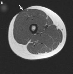
Insulin injection lipodystrophy

Many patients whit type 1 diabetes mellitus may presented with depressed areas of skin at the sites of insulin injection due to lipoatrophy......!!
Lipodystrophy or lipoatrophy is a recognized complication of Insulin injection due to loss of the adipose tissue layer at the sites of injection (prevalence of 3.6%).
Axial magnetic resonance imaging of the right thigh showed Lipoatrophy of a 14-year-old girl with a 20-month history of type 1 diabetes mellitus

The pathogenesis remains unknown, but possible mechanisms include immune reaction to insulin or excipients of the injection solution, injury from cold insulin, or trauma from repeated local injections.

Popular Posts