Custom Search
Showing posts with label ENT. Show all posts

malignant external otitis

 A 72-year-old patient withlong-standing Type 2 diabetes mellitus presents with complaints of pain in his right ear with purulent drainage. On physical exam, the patient is afebrile. The pinna of the left ear is tender, and the external auditory canal is swollen and edematous. The peripheral white blood cell count is normal. The organism most likely to grow from the purulent drainage is :
  • a. Pseudomonas aeruginosa
  • b. Staphylococcus aureus
  • c. Candida albicans
  • d. Haemophilus influenzae
  • e. Moraxella catarrhalis

 The answer is a.
Ear pain and drainage in an elderly diabetic patient must raise concern about malignant external otitis. Presence of swelling and inflammation of the external auditory meatus strongly suggests this diagnosis. This infection usually occurs in older diabetics and is almost always caused by organism Pseudomonas aeruginosa.

Haemophilus influenzae and Moraxella catarrhalis frequently cause otitis media but not external otitis.

Acute otitis externa with the canal somewhat narrowed from edema and obstructed by desquamating epithelium, soft cerumen, and purulent discharge; this must be removed to visualize the tympanic membrane and to allow ototopical therapy to penetrate to all the superficially infected areas of the canal skin.

Proposed decision tree for the management of epiglottitis

The main consideration in management is airway maintenance. Patients without signs and symptoms of airway obstruction can be treated medically in a hospital unit with equipments and personnel available for airway management if required.
A second or third generation cephalosporin is the most effective antibiotic against beta lactamase producing organisms and should be considered as initial antibiotic therapy. Simultaneous treatment of the underlying condition is mandatory. Corticisteroids have not proven in a prospective randomized trial to reduce the need for airway intervention or hasten recovery in adult acute epiglottitis.
This algorithm shows the proposed decision tree for the management of epiglottitis.
 Click her for enlargement

Picture and Imaging of Nasopharyngeal Cyst


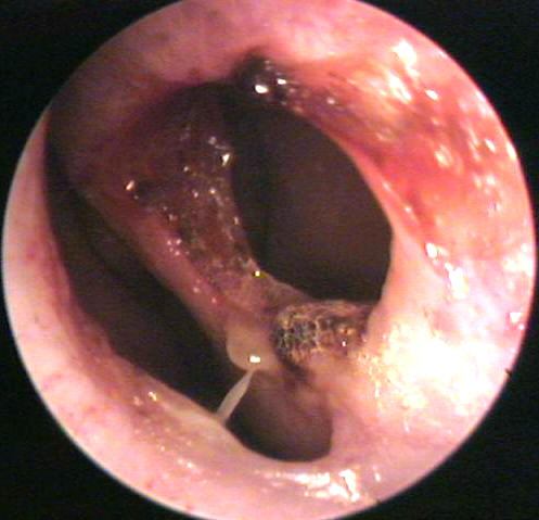
This nasopharyngeal mass on the left side was discovered during a routine nasopharyngoscopy in a 38 year-old smoker who presented with hemoptysis and epistaxis.

The CT scan below showed a mass arising from the left lateral nasopharyngeal wall. The mass was surgically removed. It turned out to be a benign mucocele.

Myringotomy tube insertion

Example of myringotomy and tube placement. Tubes are inserted for chronic are signifcant recurrent ear infections. They typically stay several months to a couple of years. They are intended to ventilate the middle ear space in order to treat as well as prevent future middle ear infections.

Photo illustrations of Bullous Myringitis

What is Bullous Myringitis ?
Bullous myringitis is painful inflammation blisters of the tympanic membrane and surrounding deep canal skin with the formation of serum or blood filled bullae. Petechial subcutaneous hemorrhages around the base of the bulla are characteristic.

Bullous Myringitis was thought that Mycoplasma pnumoniae was the most common pathogen but, recent studies have demonstrated that the pathogens are similar to otitis media. Bacteria are responsible for most cases of bullous myringitis. Streptococcus pneumoniae is the most common, followed by Haemophilus influenzae and Moraxella catarrhalis.

Examination shows a vesicular or bullous eruption over the tympanic membrane and adjacent bony canal wall. In the early stages, the vesicles are erythematous and surrounded by injected epithelium. In the later stages, they are larger and filled with fluid which may be clear and serous or blood filled. In this photograph, the bulla has now been incised with a myringotomy knife and the fluid drained. Notice the petechial hemorrhage around the base of the bulla and extending into the attic area.
Treatment of bullous myringitis requires strong analgesics because of the associated pain. Rupturing the bullae for relief of pain is controversial. Topical antibiotic eardrops are probably useful in preventing the development of a secondary bacterial otitis externa. The use of erythromycin has been advocated by those who believe that Mycoplasma is the prime etiologic agent. The tympanic membrane in this case is diffusely inflamed, with the presence of petechial hemorrhage over the attic and an associated hemotympanum


A large bulla filled with serous fluid has developed on the superficial surface of the tympanic membrane in the region of the umbo

Techniques of Ossiculoplasty - Incus Transposition

Aftere elevating tympanomeatal flap, drilling of attic area was done. Remnant incus removed. With a diamond burr and acetabulum made in the body which accomodates head of stapes.
underlaying of the temporalis fascia graft done.

Perforation of Nasal Septum


Etiology of nasal septal perforations can be classified into these 4 main categories: traumatic, iatrogenic, inflammatory/malignant, and cocaine use.

Most traumatic or iatrogenic perforations result from mucosal lacerations on corresponding sides of the septum with exposure of the underlying cartilage or from a fracture of the cartilaginous septum. Perforation occurs because the cartilage relies upon the overlying mucoperichondrium for its blood supply and nutrients.
Iatrogenic causes include nasal surgical procedures (prior septal surgery is the most common cause of septal perforations) and nasal intubation or nasogastric tube placement.

Septal hematoma, if not identified and treated early, also may result in perforation.
Infectious and inflammatory etiologies, including tuberculosis, syphilis, Wegener granulomatosis, and sarcoidosis, always should be considered in the differential diagnosis.

Treatment involves the use of nasal wetting agents, placement of a plastic button over the perforation, and surgical repair. Repair with surgery is very difficult and failure to close the perforation is common.

Granuloma of the External Auditory Canal

Granuloma defined as  highly vascular mass of fibrous tissue and blood vessels which the body forms in response to a chronic infection or healing process .
Granulomas are often seen in the ear canals in children with ear tubes. They are a result of the body attempting to heal and extrude the ear tube. Often the granuloma forms after the inner mucosal layer heals behind the eardrum then grows out the ear tube.

Treatment is steroid containing antibiotic ear drops with surgical excision reserved for persistent disease.

Facial Nerve Decompression

Young man with facial nerve paralysis and ear discharge with normal hearing.
First incudostapedial joint dislocated and modified radical mastoidectomy done.
Facial ridge has been removed. Incus removed, Chrodatympani sacrificed. Bone over the facial nerve removed. Decompression done from geniculate ganglion upto stylomastoid foramen.

Keloids of the Auricle


Keloid formation of the auricle's ear lobe is a rare complication of ear ring use. It occurs more commonly in African Americans than Caucasians. Treatments is surgical excision followed by repeated steroid injections.

Microtia ear reconstruction

This ear reconstruction video shows a surgery to correct microtia by Toronto Plastic surgeon, Leila Kasrai MD. Microtia means small ear, and it is a congenital abnormality which causes problems with the formation of the ear tissue. In some children, a small portion of tissue is seen where the ear would normally be found. In others, the ear lobe or hollow of the ear, may be partially formed with the rest of the ear tissue missing. The most severe form of this condition leaves almost no ear tissue visible.

Anatomy of ear drum as seen on examination

Normal eardrum : Notice the light reflex on the anterior-inferior portion of the eardrum.

chondrodermatitis Nodularis Helicis

This picture shows the ear of a 74 year old female who has a one year history of a slightly painful lesion on her auricle. The lesion was characteristic of chrondrodermatitis nodularis helicis (CDNH).
CDNH is a slow growing painful lesion which may reach a size of 2 to 4 mm. It begins as a nodule over the cartilage folds of the ear and may progress to form a central crust and ulceration. The cause is unknown but CDNH usually occurs on the side the patient sleeps and may be related to trauma. The most common location is on the mid portion of the antihelix.

Treatment is to excise the lesion and underlying inflamed cartilage. Steroid injections have also been advocated but in my experience they have not been successful.

Fantastic Video about Auditory Transduction

This 7-minute video by Brandon Pletsch takes viewers on a step-by-step voyage through the inside of the ear, to the acoustic accompaniment of classical music.

Exostosis in external auditory canal

Exostosis: The 1st Picture demonstrates bony ingrowths which have encroached upon the external auditory canal. Normal ear is shown for comparisno.


An osteochondroma or exostosis is a bony projection of the surface of a bone capped by cartilage.

It is the second most common benign primary bone tumour after non-ossifying fibromata.

This condition affects males and females equally in their first decade, and usually found in the lower femur, upper tibia and upper humerus i.e. the metaphysis of long bone.

The condition can be single or multiple, the later is associated with a 20% rate of malignancy, compared to less than 1% in the presence of a solitary tumour.

Popular Posts