Custom Search
Showing posts with label CLINICAL EXAMINATION: GENERAL. Show all posts

How to differentiate between transmitted and expansile pulsations

If you are palpating a swelling like an abdominal swelling infront of the aorta, You have to decide whether the mass you feel is pulsatile/expansile in itself (in which case your fingers will move outwards A ) or whether the pulsation is transmitted through other tissue (in which case your fingers will move upwards B ).See diagram below

Also in transmitted pulsation you can make this pulsation disappear if you can move the swelling away from the aorta ( if you put the patient in the knee-elbow position, an enlarged intra-abdominal swelling which was transmitting aortic pulsation will get away from the aorta and the pulsations will disappear)

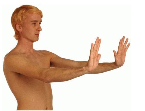
How to test Hepatic flap (asterixis)

Ask the patient to stretch out their hands in front of them with the hands dorsiflexed at the wrists and fingers outstretched and separated (see the fig.below).

The patient should hold that position for at least 15 seconds. If flap is present, the patient's hands will move in jerky, irregular flexion/extension at the wrist and MCP joints. The flap is nearly always bilateral. May be subtle and intermittent.
This is characteristic of encephalopathy due to liver failure.
If a sign of hepatic encephalopathy in a patient with previously compensated liver disease, it may have been precipitated by infection, diuretic medication, electrolyte imbalance, diarrhoea or constipation, vomiting, centrally acting drugs, upper GI bleeding, abdominal paracentesis, or surgery.
Source :oxford handbook of clinical examination

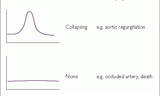
Different arterial pulse waveforms and example causes

This is best assessed at the carotid artery. You are feeling for the speed at which the artery expands and collapses and force with which it does so. It takes some practise to master and it may be useful to imagine a graph such as those shown in the figure below,also Some examples are present:

Aortic stenosis: a slow rising pulse, maybe with a palpable shudder. Sometimes called anacrotic or a plateau phase.
Aortic regurgitation: a collapsing pulse which feels as though it suddenly hits your fingers and falls away just as quickly. You could try feeling at the brachial artery and raising the arm above the patient's heart. Sometimes referred to as a waterhammer pulse.
Pulsus bisferiens: a waveform with 2 peaks, found where aortic stenosis and regurgitation co-exist.
Hypertrophic cardiomyopathy: this pulse may feel normal at first but peter out quickly. Often described as jerky.
Pulsus alternans: an alternating strong and weak pulsation, synonymous with a severely impaired left ventricle in a failing heart.
Pulsus paradoxus: pulse is weaker during inspiration (causes include cardiac tamponade, status asthmaticus, and constrictive pericarditis).


Graphical representation of different arterial pulse waveforms and their causes

Inspecting a skin lesion

 # Inspect each lesion carefully and note¦
-Grouped or solitary? Pattern if grouped .
-Distribution/location:
  • Symmetrical/asymmetrical?
  • Peripheral?
  • In only light exposed areas?
  • Dermatomal?
-Colour.
-Shape.
-Size.
-Surface.
-Edge.
-Nature of the surrounding skin.

Gynecomastia Versus Pseudogynecomastia

Gynecomastia is defined as a benign enlargement of the male breast resulting from a proliferation of the glandular component of the breast.Gynecomastia can clinically be detected by the presence of a rubbery or firm mass extending concentrically from the nipples.
Although gynecomastia is usually bilateral, it can be unilateral.

Pseudogynecomastia ( also called lipomastia ) is characterized by fat deposition without glandular proliferation.

TO SEE appearance of Pseudogynecomastia in x-ray


You can differentiate between true gynecomastia and pseudogynecomastia by a simple test made clinically by having the patient lie on his back with his hands behind his head. The examiner doctor then places a thumb on each side of the breast, and slowly brings the thumbs together. In true gynecomastia, a ridge of glandular tissue will be felt that is symmetrical to the nipple-areolar complex. With pseudogynecomastia, the fingers won't meet until they reach the nipple.
You can click on image to enlarge

Ellena's Neurological Exam


Video illustration shows neurological examination of infant reflexes.

Clubbing is Clubbing causes


It is described clinically as a bulbous uniform swelling of the soft tissue of the terminal phalanx of a digit with subsequent loss of the normal angle between the nail and the nail bed.

Since Hippocrates first described digital clubbing in patients with empyema, digital clubbing has been associated with various underlying pulmonary, cardiovascular, neoplastic, infectious, hepatobiliary, mediastinal, endocrine, and gastrointestinal diseases. Finger clubbing also may occur, without evident underlying disease


CLUBBING CAUSES:..................CLUBBING
  • Cyanotic heart disease
  • Lung disease (hypoxia, lung cancer, bronchiectasis, cystic fibrosis)
  • UC/Crohn's disease
  • Biliary cirrhosis
  • Birth defect (harmless)
  • Infective endocarditis
  • Neoplasm (esp. Hodgkins)
  • GI malabsorption

External Jugular Vein Distention


External Jugular Vein Distention:The picture above demonstrates a markedly distended right external jugular vein (EJV). This is the result of elevated central venous pressure (CVP). The picture below demonstrates the effect of hepatic pressure on the appearance of the EJV (i.e. vein becomes more engorged).
In practice the EJV is not as reliable in determining CVP as the internal jugular vein(not visualized here) due to the fact that it sometimes has valves and is not in a direct line with the right atrium. Pressure on the liver, however, will have similar impact on the appearance of the IJV as demonstrated above for the EJV. This is referred to as hepatojugular reflux.

Head and Neck Lymph nodes Exam


Lymph Nodes: The major lymph node groups are located along the anterior and posterior aspects of the neck and on the underside of the jaw. If the nodes are quite big, you may be able to see them bulging under the skin, particularly if the enlargement is asymmetric (i.e. it will be more obvious if one side is larger then the other). To palpate, use the pads of all four fingertips as these are the most sensitive parts of your hands. Examine both sides of the head simultaneously, walking your fingers down the area in question while applying steady, gentle pressure. The major groups of lymph nodes as well as the structures that they drain, are listed below. The description of drainage pathways are rough approximations as there is frequently a fair amount of variability and overlap. Nodes are generally examined in the following order:

1. Anterior Cervical (both superficial and deep): Nodes that lie both on top of
and beneath the sternocleidomastoid muscles (SCM) on either side of the neck,
from the angle of the jaw to the top of the clavicle. This muscle allows the
head to turn to the right and left. The right SCM turns the head to the left
and vice versa. They can be easily identified by asking the patient to turn
their head into your hand while you provide resistance. Drainage: The internal
structures of the throat as well as part of the posterior pharynx, tonsils,
and thyroid gland.

2. Posterior Cervical: Extend in a line posterior to the SCMs but in front of the
trapezius, from the level of the mastoid bone to the clavicle. Drainage: The
skin on the back of the head. Also frequently enlarged during upper
respiratory infections (e.g. mononucleosis).
3. Tonsillar: Located just below the angle of the mandible. Drainage: The
tonsilar and posterior pharyngeal regions.
4. Sub-Mandibular: Along the underside of the jaw on either side. Drainage: The
structures in the floor of the mouth.
5. Sub-Mental: Just below the chin. Drainage: The teeth and intra-oral cavity.
6. Supra-clavicular: In the hollow above the clavicle, just lateral to where it
joins the sternum. Drainage: Part of the throacic cavity, abdomen.

WATH THE EXAM VIDEO :

Popular Posts