
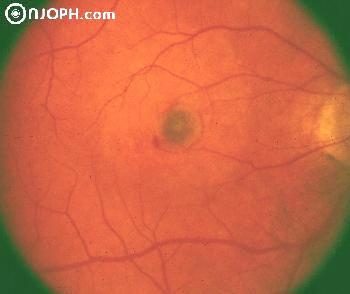
 This photo shows age-related Maculopathy AMD, Classic Choroidal Neovasculariz.
Note that in an area of slight depigmentation (presumably a previous RPE detachment) is a darkly pigmented spot surrounded by a slim ring of subretinal blood.
With Angiogram:
 #In the arterial phase the cartwheel of new choroidal vessels becomes immediately visible (together with the choroidal flush). A slight window defect is in the area of hypopigmentation.
 #In mid-angiogram the neovascularization becomes prominent. The ring of surrounding blood blocks fluorescence.
 #Late angiogram with diffuse leakage from the new vessels even into some of the blood.


