Custom Search
Showing posts with label ANATOMY. Show all posts

How to remember Carpal bones !!

"She Looks Too Pretty; Try To Catch Her":
· Proximal row then distal row, both lateral-to-medial:
  • Scaphoid
  • Lunate
  • Triquetrium
  • Pisiform
  • Trapezium
  • Trapezoid
  • Capate
  • Hamate
· Alternatively: "She Likes To Play; Try To Catch Her".

Note :The scaphoid bone is a small carpal bone on the thumb side (radial side) of the wrist. It is the most commonly fractured carpal bone.

This is probably because it actually crosses two rows of carpal bones, forming a hinge.

Surface anatomy of both kidneys on the back

Viewed posteriorly the right kidney has its upper edge opposite the 11th dorsal spine and the lower edge of the 11th rib. Its lower edge is opposite the upper edges of L3 spine and vertebral body and about 4 cm. (1 1/2 in.) above the highest point of the crest of the ileum, which is opposite the fourth spine.The left kidney is usually 1.25 cm. (1/2 in.) higher, but being a little longer than the right, its lower limit may not be quite that much higher. The kidney is slightly lower in women and children than in men. The inner border reaches 10 cm. (4 in. ) and the hilum 4 to 5 cm. (1 1/2 to 2 in.)
Notice that the left kidney is higher (highest border T11 lowest border L2) whereas the right kidney is lower (highest border T12 lowest border L3). The right kidney is less enclosed by the rib cage, because of the presence of right lobe of liver above it, therefore pushing it down. So, the right kidney is palpable during inspiration (moves down by 1 inch).

The renal angle as seen in the image, is located between the

-lower border of the 12th rib
-& lateral border of the erector spinae muscle
Left kidney related to 11th & 12th rib. Right kidney related to only 12th rib.

 Also to indicate the position of the kidney from the back, the parallellogram of Morris is used; two vertical lines are drawn, the first 2.5 cm., the second 9.5 cm. from the middle line; the parallelogram is completed by two horizontal lines drawn respectively at the levels of the tips of the spinous process of the eleventh thoracic and the lower border of the spinous process of the third lumbar vertebra. The hilum is 5 cm. from the middle line at the level of the spinous process of the first lumbar vertebra.
  Back of lumbar region, showing surface markings for kidneys, ureters, and spleen. The lower portions of the lung and pleura are shown on the right side.

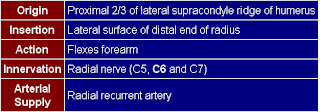
B.R. of BrachioRadialis ;Medical mnemonic

BrachioRadialis:
Function: Its the Beer Raising muscle, flexes elbow, strongest when wrist is oriented like holding a beer.
Innervation: Breaks Rule: it's a flexor muscle, But Radial. (Radial nerve usually is for extensors: Recall BEST rule: B was for brachioradialis).
Important relation: Behind it is the Radial nerve in the cubital fossa.
Attachment: Attaches to Bottom of Radius.

Anatomy of spinal nerves exiting through the intervertebral foramina

The spinal cord is an extension of the brainstem (the lowest part of the brain), extending from the base of the skull down to the low back. Along its length, it gives off 31 pairs of spinal nerves, which branch to form the peripheral nerves of the neck, trunk, and extremities. The origins of the spinal nerves as they emerge from the spinal cord are called nerve roots.


This image shows a dissection of the cervical region, showing a posterior view of cervical spinal nerves exiting the intervertebral foramina on the right side.


The spinal nerves exit from the vertebral column through openings between adjacent vertebrae. These openings, called intervertebral foramina, are located just in front of the facet joints. The spinal nerves are named and numbered according to the vertebral levels at which they exit. There are eight paired cervical nerves (C1-C8), twelve thoracic (T1-T12), five lumbar (L1-L5), five sacral (S1-S5), and one coccygeal (Co1).
Note :
-There are eight pairs of cervical nerves, although there are only seven cervical vertebrae. The C1 nerves exit above the C1 vertebra (between C1 and the base of the skull).
-Although the spinal nerves correspond to their respective vertebral levels, the spinal cord itself is shorter than the vertebral column, extending only as far as the L1 vertebra. Below that level, the spinal canal contains only the roots of the lumbar, sacral, and coccygeal nerves, as they descend to exit at the appropriate levels. This bundle of descending nerve roots is called the cauda equina (Latin for "horse's tail").


The on the right views a portion of the spinal cord, showing its right lateral surface. The dura is opened and arranged to show the nerve roots.


Nerve Roots:—
Each nerve is attached to the medulla spinalis by two roots, an anterior = ventral, and a posterior = dorsal, the latter being characterized by the presence of a ganglion, the spinal ganglion.

Blood and nerve supply of a Synovial joint

The blood supply of a synovial joint is derived from the arteries sharing in the anastomosis around the joint.And the nerve supply of a synovial joint is derived from the muscles which act on the joint ,best expressed by Hilton in 1863:
"The same trunks of nerves, whose branches supply the groups of muscles moving a joint, furnish also a distribution of nerves to the skin over the insertions of the same muscles; and what at this moment more especially merits our attention-the interior of the joint receives its nerves from the same source."

proprioceptive fibers endings in the capsule and ligaments are very sensitive to position and movement. Their central connections are such that they are concerned with the reflex control of posture and locomotion and the detection of position and movement.

The blood and nerve supply of a synovial joint. This diagram shows the artery supplying the epiphysis, joint capsule, and synovial membrane. The nerve that contains (1) sensory (mostly pain) fibers from the capsule and synovial membrane, (2) autonomic (postganglionic sympathetic) fibers to blood vessels, (3) sensory (pain) fibers from the adventitia of blood vessels, and (4) proprioceptive fibers. 
N.B.Arrowheads indicate direction of conduction.

Diagram of Anterior anatomical relations of both kidneys

The kidneys are retroperitoneal organs that are located in the perirenal retroperitoneal space with a longitudinal diameter of 10–12 cm and a latero-lateral diameter of 3–5 cm and a weight of 250–270 g.
In the supine position, the medial border of the normal kidney is much more anterior than the lateral border, The upper pole of each kidney is situated more posteriorly than the lower pole.
The right kidney,  anteriorly :
has a relation with the inferior surface of the liver with peritoneal interposition,and with the second portion of the duodenum without any peritoneal interposition since the second portion of the duodenum is retroperitoneal .
The left kidney, anteriorly :
has a relation with the pancreatic tail, the spleen, the stomach, the ligament of Treitz and small bowel, and with the left colic lexure and left colon . Over the left kidney, there are two important peritoneal relections, one vertical corresponding to the spleno-renal ligament (connected to
the gastro-diaphragmatic and gastrosplenic ligaments) and one horizontal corresponding to the transverse mesocolon.

Good Mnemonics for Thenar and hypothenar muscles

FAO (Flex, Oppose, Abduct)

Thenar:
Flexor pollicis brevis, Opponens pollicis, Abductor pollicis brevis.


Hypothenar:
Flexor digiti minimi, Abductor digiti minimi, Opponens digiti minimi.

Arterial supply of the Stomach

The arteries that supply the stomach are branches of the celiac trunk . This is the first unpaired branch of the abdominal aorta, arising just after the aorta passes behind the diaphragm.
The branches of the celiac artery are three:

1. left gastric
2. splenic
3. common hepatic

* celiac "C"
o left gastric LG - supplies the lesser curvature of the stomach and lower esophagus
+ esophageal E
* splenic "S" which gives rise to:
o short gastric SG - supplies area of the fundus
o left gastroepiploic LGE - supplies the left part of greater curvature of the stomach
* common hepatic "CH"
o gastroduodenal GD
+ right gastric RG - supplies right side of lesser curvature of the stomach
+ right gastroepiploic RGE - supplies the right part of the greater curvature of the stomach

Action of lateral vs. medial Pterygoid muscle "mnemonic"

Look at how your jaw ends up when saying first syllable of 'Lateral' or 'Medial' :
  1. -"La": your jaw is now open, so Lateral opens mouth.
  2. -"Me": your jaw is still closed, so medial closes the mandible.

medial pterygoid muscle
Origin : deep head: medial side of lateral pterygoid plate behind the upper teeth
superficial head: pyramidal process of palatine bone and maxillary tuberosity
Insertion : medial angle of the mandible
Actions : elevates mandible, closes jaw, helps lateral pterygoids in moving the jaw from side to side

Lateral pterygoid muscle
Origin :  Great wing of sphenoid and pterygoid plate
Insertion : Condyle of mandible
Actions : depresses mandible, Protrude mandible, side to side movement of mandible

Terms of Position, Direction and the main Planes of human body anatomy

The Diagram below shows the chief terms of position and direction and the main planes of reference in the body.
*A convention whereby the body is erect, with the head, eyes, and toes directed forward and the upper limbs by the side and held so that the palms of the hands face forward "unlike the figure at left " . It is often necessary, however, to describe the position of the viscera also in the recumbent posture, because this is a posture in which patients are frequently examined clinically. 

*The median plane is an imaginary vertical plane of section that passes longitudinally through the body and divides it into right and left halves. The median plane intersects the surface of the front and back of the body at what are called the anterior and posterior median lines. It is a common error, however, to refer to the" midline" when the median plane is meant.

*Any vertical plane through the body that is parallel with the median plane is called a sagittal plane. The sagittal planes are named after the sagittal suture of the skull, to which they are parallel. The term "parasagittal" is redundant: anything parallel with a sagittal plane is still sagittal.

*The term horizontal plane refers to a plane at a right angle to both the median and coronal planes: it separates the body into superior and inferior parts. This is often termed an axial plane, particularly in radiology.

*The term transverse means at a right angle to the longitudinal axis of a structure. Thus, a transverse section through an artery is not necessarily horizontal. A transverse section through the hand is horizontal, whereas a transverse section through the foot is coronal !!

*The suffix "-ad" is sometimes added to a positional term to indicate the idea of motion. Thus, cephalad means proceeding toward the head. Such terms are useful occasionally in describing growth processes, but their application is best limited.

Layers of the Abdominal Wall with Function

The layers of the abdominal wall vary, depending on where it is you are looking.
At the lateral side of the abdomen there is a dotted line passing through the abdominal wall. Note the layers must pass through to get to the peritoneal cavity:

1. skin
2. superficial fascia (this may be as thin as or less than a half inch or as thick as 6 inches or more)
3. deep fascia (all skeletal muscle is surrounded within its own deep fascia). The deep fascia of the abdominal wall is different than that found around muscles of the extremities, however. It is of the loose connective tissue variety. It is necessary in the abdominal wall because it offers more flexibility for a variety of functions of the abdomen. At certain points, this fascia may become aponeurotic and serve as attachments for the muscle to bone or to each other, as is the case at the linea alba.
4. subserous fascia also known at extraperitoneal fascia (a layer of loose connective tissue that serves as a glue to hold the peritoneum to the deep fascia of the abdominal wall or to the outer lining of the GI tract. It may receive different names depending on its location (i.e. transversalis fascia when it is deep to that muscle, psoas fascia when it is next to that muscles, iliac fascia, etc.)
5. peritoneum (a thin one cell thick membrane that lines the abdominal cavity and in certain places reflects inward to form a double layer of peritoneum) Double layers of peritoneum are called mesenteries, omenta, falciform ligaments, lienorenal ligament, etc.)

--At the anterior wall of the abdomen, in the midline there is no muscle.

--If we look at the wall inferior to the level of the belly button (umbilicus), you will see that the superficial fascia has become divided into 2 parts:
* a superficial fatty part that is continuous with the same layer over the rest of the body (Camper's fascia)
* a deep membranous layer that is continuous down into the perineum to surround the penis and to form a layer of the scrotum. (Scarpa's fascia)

The lady between two majors


Bicipital groove: attachments of muscles near it

"The lady between two majors" :
Teres major attaches to medial lip of groove.
Pectoralis major to lateral lip of groove.
Latissimus (Lady) is on floor of groove, between the 2 majors.

Anatomy on Thyroid sonography

The anterior neck is depicted rather well with standard gray scale sonography. (FIGURE 1) The thyroid gland is slightly more echo-dense than the adjacent structures because of its iodine content. It has a homogenous ground glass appearance. Each lobe has a smooth globular-shaped contour and is no more than 3 - 4 centimeters in height, 1 - 1.5 cm in width, and 1 centimeter in depth. The isthmus is identified, anterior to the trachea as a uniform structure that is approximately 0.5 cm in height and 2 - 3 mm in depth.
The pyramidal lobe is not seen unless it is significantly enlarged. In the female, the upper pole of each thyroid lobe may be seen at the level of the thyroid cartilage, lower in the male. The surrounding muscles are of lower echogenicity than the thyroid and tissue planes between muscles are usually identifiable. The air-filled trachea does not transmit the ultrasound and only the anterior portion of the cartilaginous ring is represented by dense, bright echoes. The carotid artery and other blood vessels are echo-free unless they are calcified. The jugular vein is usually in a collapsed condition and it distends with a Valsalva maneuver. There are frequently 1-2 mm echo-free zones on the surface and within the thyroid gland that represent blood vessels. The vascular nature of all of these echoless areas can be demonstrated by color Doppler imaging to differentiate them from cystic structures (10-11).
Lymph nodes may be observed and nerves are generally not seen. The parathyroid glands are observed only when they are enlarged and are less dense ultrasonically than thyroid tissue because of the absence of iodine. The esophagus may be demonstrated behind the medial part of the left thyroid lobe, especially if it is distended by a sip of water. (FIGURE 2)
Figure 1. Sonogram of the neck in the transverse plane showing a normal right thyroid lobe and isthmus. L=small thyroid lobe in a patient who is taking suppressive amounts of L-thyroxine, I=isthmus, T=tracheal ring ( dense white arc is calcification, distal to it is artefact), C=carotid artery ( note the enhanced echoes deep to the fluid-filled blood vessel), J=jugular vein, S=Sternocleidomastoid muscle, m=strap muscle.

Figure 2. Sonogram of the left lobe of the thyroid gland in the transverse plane showing a rounded lobe of a goiter. L=enlarged lobe, I= widened isthmus, T=trachea, C=carotid artery ( note the enhanced echoes deep to the fluid-filled blood vessel), J=jugular vein, S=Sternocleidomastoid muscle, m=strap muscles, E=esophagus.

Trachea and Esophagus in relation to vertebral and sternal levels

The trachea, or windpipe , which has cervical and thoracic parts, extends from the inferior end of the larynx (C6 vertebra) to its point of bifurcation (between T5 and 7 vertebral level). It is about 9 to 15 cm in length. The trachea descends anterior to the esophagus, enters the superior mediastinum, and divides into right and left main bronchi. The trachea is a median structure but, near its lower end, deviates slightly to the right, resulting in the left main bronchus crossing anterior to the esophagus. Owing to the translucency of the air within it, the trachea is usually visible above the arch of the aorta in radiographs.

The trachea has 15 to 20 C-shaped bars of hyaline cartilage that prevent it from collapsing. Longitudinal elastic fibers enable the trachea to stretch and descend with the roots of the lungs during inspiration. When a subject is in the erect position, the trachea divides between the T5 and T7 vertebral levels. The carina is the upward-directed ridge seen internally at the bifurcation and is a landmark during bronchoscopy.
The trachea and esophagus in relation to vertebral and sternal levels in a subject in the erect position.

The arch of the aorta is at first anterior to the trachea and then on its left side immediately superior to the left main bronchus. Other close relations include the brachiocephalic and left common carotid arteries. The trachea is supplied mainly by the inferior thyroid arteries. Its smooth muscle is supplied by parasympathetic and sympathetic fibers, and pain fibers are carried by the vagi.

Popular Posts