Recession, Limited Gingival Band, Esthetics and Stability are addressed with this Periodontal Microsurgical procedure using a purified collagen implant: AlloDerm

Custom Search
smooth-muscle cells compared to skeletal muscle
Which of the following is absent in smooth-muscle cells compared to skeletal muscle cells?
The answer is (a).
Smooth muscle is the least specialized type of muscle and contains no troponin. The contractile process is similar to the actin-myosin interactions that occur in motility of nonmuscle cells. In the smooth-muscle cell, actin and myosin are attached to intermediate filaments at dense bodies in the sarcolemma and cytoplasm. Dense bodies contain alpha-actinin and, therefore, resemble the Z-lines of skeletal muscle. Contraction causes cell shortening and a change in shape from elongate to globular. Contraction occurs by a sliding filament action analogous to the mechanism used by thick and thin filaments in striated muscle.
 The connections to the plasma membrane allow all the smooth-muscle cells in the same region to act as a functional unit. The sarcoplasmic reticulum is not as well developed as that in the striated muscles. There are no T tubules present; however, endocytic vesicles called caveolae are believed to function in a fashion similar to the T tubule system of skeletal muscle. When intracellular calcium levels increase, the calcium is bound to the Ca2+ -binding protein, calmodulin. Ca2+-calmodulin (answers b and c) is required and is bound to myosin light-chain kinase (answer d) to form a Ca2+ -calmodulin-kinase complex. This complex catlayzes the phosphorylation of one of the two myosin light chains on the myosin heads. That phosphorylation allows the binding of actin to myosin. A specific phosphatase
dephosphorylates the myosin light chain, which returns the actin and myosin to the inactive, resting state. The actin-tropomyosin interactions (answer e) are similar in smooth and skeletal muscle.
Smooth-muscle cells (e.g., vascular smooth-muscle cells) also differ from skeletal muscle cells in that like fibroblasts, they are capable of collagen, elastin, and proteoglycan synthesis.
- a.Troponin
- b.Calmodulin
- c.Calcium
- d.Myosin light-chain kinase
- e.Actin and tropomyosin interactions
The answer is (a).
Smooth muscle is the least specialized type of muscle and contains no troponin. The contractile process is similar to the actin-myosin interactions that occur in motility of nonmuscle cells. In the smooth-muscle cell, actin and myosin are attached to intermediate filaments at dense bodies in the sarcolemma and cytoplasm. Dense bodies contain alpha-actinin and, therefore, resemble the Z-lines of skeletal muscle. Contraction causes cell shortening and a change in shape from elongate to globular. Contraction occurs by a sliding filament action analogous to the mechanism used by thick and thin filaments in striated muscle.

dephosphorylates the myosin light chain, which returns the actin and myosin to the inactive, resting state. The actin-tropomyosin interactions (answer e) are similar in smooth and skeletal muscle.
Smooth-muscle cells (e.g., vascular smooth-muscle cells) also differ from skeletal muscle cells in that like fibroblasts, they are capable of collagen, elastin, and proteoglycan synthesis.
Good Mnemonics for Thenar and hypothenar muscles
FAO (Flex, Oppose, Abduct)
Thenar:
Flexor pollicis brevis, Opponens pollicis, Abductor pollicis brevis.
Hypothenar:
Flexor digiti minimi, Abductor digiti minimi, Opponens digiti minimi.

Thenar:
Flexor pollicis brevis, Opponens pollicis, Abductor pollicis brevis.
Hypothenar:
Flexor digiti minimi, Abductor digiti minimi, Opponens digiti minimi.

Acute Upper Airway Obstruction (Epiglottitis Vs Croup)
Most children who present to the hospital with acute upper airway obstruction have croup (acute laryngo- tracheobronchitis) or epiglottitis. Other differential diagnoses include bacterial tracheitis, foreign body inhalation and angioneurotic oedema.
You have to assess severity by examination of
Guidelines For The Diagnosis Of Croup And Epiglottitis:

2. If the child is cyanosed he is likely to die very quickly -notify paediatric ICU immediately. The aim is to transfer severely obstructed children to ICU before intubation becomes imperative, as it is preferable to intubate them in ICU.
3. as a child becomes physically exhausted the stridor, indrawing and air entry may decrease.
You have to assess severity by examination of
- constitutional state (toxicity, fever, pulse rate)
- stridor
- drooling
- cough
- speech
- tachypnoea
- tracheal tug on inspiration
- intercostal and subcostal indrawing on inspiration
- asynchrony of chest and abdominal wall movement
- cyanosis in air
Guidelines For The Diagnosis Of Croup And Epiglottitis:

Note: Each of these clinical signs in isolation is a poor discriminator however, considered together, they usually allow the correct diagnosis to be made.
Important Notes
1. All children with stridor must be reviewed by the paediatric admitting officer before discharge home or admission to the ward.2. If the child is cyanosed he is likely to die very quickly -notify paediatric ICU immediately. The aim is to transfer severely obstructed children to ICU before intubation becomes imperative, as it is preferable to intubate them in ICU.
3. as a child becomes physically exhausted the stridor, indrawing and air entry may decrease.
Mallet finger :Injury to the extensor tendon at the DIP joint
Injury to the extensor tendon at the DIP joint is also known as :
 Mallet finger usually is caused by an object (e.g., a ball ;baseball finger) striking the finger, creating a forced flexion of an extended DIP. The extensor tendon may be strained, partially torn, or completely ruptured or separated by a distal phalanx avulsion fracture. Those affected with mallet finger complain of pain at the dorsal DIP joint; inability to actively extend the joint; and, often, with a characteristic flexion deformity. It is critical to isolate the DIP joint during the evaluation to ensure extension is from the extensor tendon and not the central slip. The absence of full passive extension may indicate bony or soft tissue entrapment requiring surgical intervention.

- A) boutonnière deformity
- B) jersey finger
- C) mallet finger
- D) swan necking
- E) “jammed” finger
The answer is (C).
Injury to the extensor tendon at the DIP joint is also known as Mallet Finger or Dropped Finger. The condition is the most common closed tendon injury of the finger.
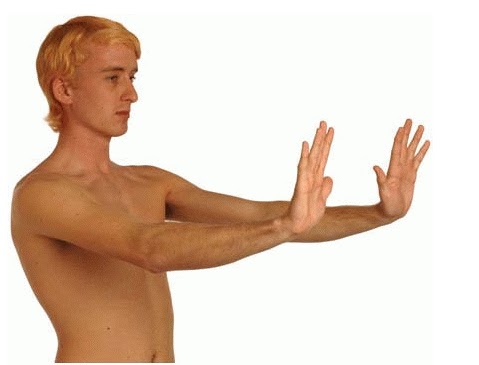
How to test Hepatic flap (asterixis)
Ask the patient to stretch out their hands in front of them with the hands dorsiflexed at the wrists and fingers outstretched and separated (see the fig.below).

The patient should hold that position for at least 15 seconds. If flap is present, the patient's hands will move in jerky, irregular flexion/extension at the wrist and MCP joints. The flap is nearly always bilateral. May be subtle and intermittent.
This is characteristic of encephalopathy due to liver failure.
If a sign of hepatic encephalopathy in a patient with previously compensated liver disease, it may have been precipitated by infection, diuretic medication, electrolyte imbalance, diarrhoea or constipation, vomiting, centrally acting drugs, upper GI bleeding, abdominal paracentesis, or surgery.
Source :oxford handbook of clinical examination
Innate immunity Vs Adaptive immunity with Brief Video Review
Immune System has 2 arms:
 I
I
Innate immunity (natural or native immunity)
Instant
Immediate
Initial response
Induces adaptive immunity
Integrates with adaptive immunity
Acquired
Await days = no immediate response
Accurate = specific
Autoregulation
Autoimmunity
Innate immunity is the first line of defense against infections. Innate immunity is specifically against microbes, while adaptive immunity is against any "foreign" substance (including cancer cells, autoantigens, etc.).
Innate immunity is the oldest mechanism of defense. Adaptive immunity (T and B lymphocytes) appeared in jawed vertebrates (sharks) and is superimposed on innate immunity to improve host defense. In a sense, adaptive immunity is an "add-on" to innate immunity.
- - Innate immune system
- - Adaptive immune system

Innate immunity (natural or native immunity)
Instant
Immediate
Initial response
Induces adaptive immunity
Integrates with adaptive immunity
A
Adaptive immune systemAcquired
Await days = no immediate response
Accurate = specific
Autoregulation
Autoimmunity
The Immune Response
Innate immunity is the first line of defense against infections. Innate immunity is specifically against microbes, while adaptive immunity is against any "foreign" substance (including cancer cells, autoantigens, etc.).
Innate immunity is the oldest mechanism of defense. Adaptive immunity (T and B lymphocytes) appeared in jawed vertebrates (sharks) and is superimposed on innate immunity to improve host defense. In a sense, adaptive immunity is an "add-on" to innate immunity.
Innate Recognition of Pathogens
Repair of Tetralogy of Fallot with Atrioventricular Canal
Redmond Burke MD, Chief of Pediatric Cardiovascular Surgery at Miami Children's Hospital demonstrates the repair of TOF with AVC, Tetralogy of Fallot with Atrioventricular Canal. The video is graphic, showing the operative repair and the postoperative recovery. The program website is www.pediatricheartsurgery.com
Granuloma of the External Auditory Canal

Granulomas are often seen in the ear canals in children with ear tubes. They are a result of the body attempting to heal and extrude the ear tube. Often the granuloma forms after the inner mucosal layer heals behind the eardrum then grows out the ear tube.
Treatment is steroid containing antibiotic ear drops with surgical excision reserved for persistent disease.
iron deficiency anemia in pregnancy
A 22-year-old woman is pregnant and at 14-week gestation. Her hemoglobin level is 9 g/dL. She asks why she could have iron deficiency when she is no longer menstruating. Which of the following is the best explanation?
 ( B ). Iron deficiency occurs in pregnancy as a result of the expanded blood volume and active transport of iron to the fetus.
- A.Occult gastrointestinal blood loss
- B.Expanded blood volume and transport to the fetus
- C.Hemolysis
- D.Iron losses as a result of relative alkalosis of pregnancy
Facial Nerve Decompression
Young man with facial nerve paralysis and ear discharge with normal hearing.
First incudostapedial joint dislocated and modified radical mastoidectomy done.
Facial ridge has been removed. Incus removed, Chrodatympani sacrificed. Bone over the facial nerve removed. Decompression done from geniculate ganglion upto stylomastoid foramen.
First incudostapedial joint dislocated and modified radical mastoidectomy done.
Facial ridge has been removed. Incus removed, Chrodatympani sacrificed. Bone over the facial nerve removed. Decompression done from geniculate ganglion upto stylomastoid foramen.
Arterial supply of the Stomach
The arteries that supply the stomach are branches of the celiac trunk . This is the first unpaired branch of the abdominal aorta, arising just after the aorta passes behind the diaphragm.
The branches of the celiac artery are three:
1. left gastric
2. splenic
3. common hepatic

+ esophageal E
* splenic "S" which gives rise to:
o short gastric SG - supplies area of the fundus
o left gastroepiploic LGE - supplies the left part of greater curvature of the stomach
+ right gastric RG - supplies right side of lesser curvature of the stomach
+ right gastroepiploic RGE - supplies the right part of the greater curvature of the stomach
The branches of the celiac artery are three:
1. left gastric
2. splenic
3. common hepatic

* celiac "C"
o left gastric LG - supplies the lesser curvature of the stomach and lower esophagus+ esophageal E
* splenic "S" which gives rise to:
o short gastric SG - supplies area of the fundus
o left gastroepiploic LGE - supplies the left part of greater curvature of the stomach
* common hepatic "CH"
o gastroduodenal GD+ right gastric RG - supplies right side of lesser curvature of the stomach
+ right gastroepiploic RGE - supplies the right part of the greater curvature of the stomach
Everything You Need To Know about Ankle fracture
ANKLE FRACTURE ,Educational animation video describing the anatomy the injury the diagnosis and treatment of ankle fracture
ankle fracture are single malleolar bimalleolar trimalleolar
syndesmodic injuy is diagnosed and the ankle is fixed to reduce the talus under the dome of the tibia
any lateral shift will increase the stress by 40 to 50 percent and accelerate arthritis
this ankle fracture video is uploaded by the univresity of toledo orthopedic surgeon
Picture for Klinefelter syndrome

a.The patient is likely to have low levels of gonadotropins
b.The patient has Turner syndrome
c.His most likely karyotype is 47 XXY
d.The patient will have normal sperm count and testosterone level
The answer is (c).
The picture of infertility,gynecomastia, and tall stature is consistent with Klinefelter syndrome and an XXY karyotype. The patient has abnormal gonadal development with hyalinized testes that result in low testosterone levels and elevated levels of gonadotropin. Turner syndrome refers to the 45 XO karyotype that resultsin abnormal sexual development in a female.
Frostbite

It most commonly affects areas that are further away from the body core and have less blood flow. These include your feet, hands, nose, and ears.
There are three degrees of cold injury: frostnip, superficial frostbite, and deep frostbite.

The affected skin may be slightly flushed. The skin changes to white or grayish yellow as the frostbite develops. Pain is sometimes felt early but subsides later. Often there is NO pain; the part being frostbitten simply feels intensely cold and numb.
In superficial frostbite, there will be an area that looks white or grayish and the surface skin will feel hard but the underlying tissue will be soft. With deeper involvement, large blisters appear on the surface, as well as in underlying tissue, and the affected area is hard, cold and insensitive. Destruction of the entire thickness of the skin will necessitate skin grafting and will constitute a medical emergency, because gangrene may result from loss of blood supply to the injured part.

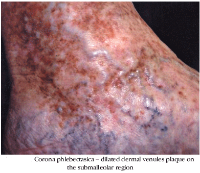
Corona phlebectatica in venous leg ulcer
Some patients with venous leg ulcer have a plaque of intradermal dilated venules, usually at the ankle, on the submalleolar region. This is known as corona phlebectatica, and results from persistent venous hypertension, leading to dilation and elongation of capillaries and venules .

Chickenpox infection
A 25 years old teacher develops fever and an itchy rash over her face and chest. on exam : multiple papules and vesicles in varying stages of development. 1 week later she complains of cough and is found to have an infiltrate on x-ray.the most likely etiology of the infection?

- a. Streptococcus pneumoniae
- b. Mycoplasma pneumoniae
- c. Pneumocystis carinii
- d. Varicella virus
- e. Chlamydia psitacii

The answer is d.
Varicella pneumonia develops in about 20% of adults with chickenpox. It occurs 3 to 7 days after the onset of the rash. The hallmark of the chickenpox rash is papules, vesicles, and scabs in various stages of development. Fever, malaise, and itching are usually part of the clinical picture. The differential can include some coxsackievirus and echovirus infections, which might present with pneumonia and vesicular rash. Rickettsialpox, a rickettsial infection, has also been mistaken for chickenpox.
lip filler (Restylane Lipp) treatment
Here is one of the patients having lip filler (Restylane Lipp) treatment on her upper lip which is performed by Tracey BellDermal Fillers are an excellent treatment option for:
Lip enhancement
Volume augmentation of the lips
Shaping facial contours (such as cheeks and chin correcting)
Smile lines (correcting thin, superficial lines around the eyes, mouth and forehead)
Lip enhancement
Volume augmentation of the lips
Shaping facial contours (such as cheeks and chin correcting)
Smile lines (correcting thin, superficial lines around the eyes, mouth and forehead)
Simple illustrations of Carotid Endarterectomy Procedure

Carotid Endarterectomy or may be called Stroke Prevention Surgery is indicated to prevent a stroke in patients with narrowed carotid arteries(Endarterectomy is the removal of material on the inside "end-" of an artery).

Procedure: The carotid artery is exposed through an incision on the side of the neck. The artery is clamped above and below where the narrowing is located. The flow of blood to the brain is maintained with the use of a specialized shunt (tube) that carries fresh blood to the brain during the procedure. The diseased material narrowing the artery is removed. The artery is then closed.


Neural transmission in the nodes of Ranvier
The nodes of Ranvier increase the efficiency of neural transmission by means of which of the following?
the space between adjacent units of myelination.
This area is bare in the CNS, whereas in the PNS the axons in the nodes are partially covered by the cytoplasmic tongues of adjacent Schwann cells. Most of the Na -gated channels are located in the bare areas. Therefore, spread of depolarization from the nodal region along the axon occurs until it reaches the next node.
This is often described as a series of jumps from node to node, or saltatory conduction.
- a.Decelerating the closing of Na -gated channels
- b.Enhancing myelination of the internodal segment
- c.Sequestration of Na entry into the axon
- d.Multiple firings due to local ionic currents around the node
- e.Decreasing threshold for the action potential
The answer is (c ).
The nodes of Ranvier increase the efficiency of nodal conduction because of restriction (sequestration) of energy–dependent Na influx to the node. The nodes of Ranvier representthe space between adjacent units of myelination.
This area is bare in the CNS, whereas in the PNS the axons in the nodes are partially covered by the cytoplasmic tongues of adjacent Schwann cells. Most of the Na -gated channels are located in the bare areas. Therefore, spread of depolarization from the nodal region along the axon occurs until it reaches the next node.
This is often described as a series of jumps from node to node, or saltatory conduction.
About anaerobic infection
A 40-year-old alcoholic develops cough and fever. Chest x-ray shows an air-fluid level in the superior segment of the right lower lobe. Which of the following is the most likely etiologic agent?
 Of the organisms listed, only anaerobic infection is likely to cause a necrotizing process. S. pneumoniae capsular type III pneumococci have been reported to cause cavitary disease, but this is unusual.
The location of the infiltrate suggests aspiration, also making anaerobic infection most likely. The superior segment of the right lower lobe is the one most likely to develop an aspiration pneumonia.
- a. Streptococcus pneumoniae
- b. Haemophilus influenzae
- c. Legionella
- d. Anaerobes
- e. Mycoplasma pneumoniae
The answer is " d ".

The location of the infiltrate suggests aspiration, also making anaerobic infection most likely. The superior segment of the right lower lobe is the one most likely to develop an aspiration pneumonia.
Diagram for possible long-term effects of ethanol
This diagram shows most significant possible long-term effects of ethanol.


Press for enlargement
Simple Terms to learn about effect of drugs on CNS

- Addiction : The state of response to a drug whereby the drug taker feels compelled to use the drug and suffers anxiety when separated from it
- Anesthesia : Loss of consciousness associated with absence of response to pain
- Anxiolytic : A drug that reduces anxiety, a sedative
- Dependence : The state of response to a drug whereby removal of the drug evokes unpleasant, possibly life-threatening symptoms, often the opposite of the drug's effects
- Hypnosis : Induction of sleep
- REM : sleep Phase of sleep associated with rapid eye movements; most dreaming takes place during REM sleep
- Tolerance : Reduction in drug effect requiring an increase in dosage to maintain the same response
Placement Percutaneous Endoscopic Gastrostomy tube
Placement of a Flocare PEG tube (Percutaneous Endoscopic Gastrostomy tube) in a patient who previously was fed via a naso-gastric tube.
The video shows detailed instructions for placement, immediate care and long-term care of the PEG tube.
The video shows detailed instructions for placement, immediate care and long-term care of the PEG tube.
Raccoon eyes ,a sign for basal skull fracture

Raccoon eyes is also known in the UK as panda eyes or periorbital ecchymosis that is a sign of basal skull fracture or a craniotomy that ruptured the meninges.And not due to facial soft-tissue trauma.
It is from bilateral subconjunctival hemorrhage, and occurs when damage at the time of fracture tears the meninges and causes the venous sinuses to bleed into the arachnoid villi and the cranial sinuses.It developes 2-3 days after a closed head injury that results in a basilar skull fracture.
Note :Somtimes Raccoon eyes are accompanied by Battle's sign (HERE)

Explanation of the state of rigor mortis

- a.Inhibition of Ca++ leakage from the extracellular fluid and sarcoplasmic reticulum
- b.Enhanced retrieval of calcium by the sarcoplasmic reticulum
- c.Failure to disengage tropomyosin and troponin from the myosin active sites
- d.Absence of ATP preventing detachment of the myosin heads from actin
- e.Increased lactic acid production
The answer is d.
There is some small amount of production of ATP after death through anaerobic and phosphagen pathways. However, there is insufficient ATP to induce the detachment of the myosin heads from actin. Ca ions continue to leak from the extracellular fluid and the sarcoplasmic reticulum (answer a),however, the sarcoplasmic reticulum is no longer able to retrieve the Ca ions (answer b). Tropomyosin and troponin are disengaged from the myosin active sites (answer c). Lactic acid is produced during rigor mortis through anaerobic pathways. The high levels of lactic acid cause deterioration of the skeletal muscle and end the state of rigor mortis (answer e).
Bilirubin metabolism made simple
A medical student teaching video on heme catabolism, bilirubin metabolism in the liver. Particularly useful in understanding the difference between direct and indirect bilirubin and why they differ in hemolytic anemias
Preferred method for chest compressions in neonatal resuscitations
In the most of neonatal resuscitations, if adequate ventilation is achieved, no need for chest compressions . However, in certain cases of advanced asphyxia and myocardial depression or severe pulmonary dysfunction in which adequate ventilation cannot be readily achieved, chest compressions are necessary to support the circulation during more extensive resuscitation.
Thus, the indication for chest compressions in the newly born differs significantly from that in older children and adults. The mechanics of the thoracic cage and the physical forces of the circulation of blood also differ, especially in preterm infants. The predominance of pulmonary dysfunction, necessitates a relatively lower ratio of compressions to ventilations. The 3:1 ratio of compressions to ventilations is performed with 90 compressions and 30 interposed breaths per minute (or one cycle of 4 events every 2 seconds).
 The preferred method for chest compressions is the two-thumb-encircling-hands method , which provides firm support for the back and generates higher systemic arterial pressure and better coronary perfusion pressure than the two-finger method.
Thus, the indication for chest compressions in the newly born differs significantly from that in older children and adults. The mechanics of the thoracic cage and the physical forces of the circulation of blood also differ, especially in preterm infants. The predominance of pulmonary dysfunction, necessitates a relatively lower ratio of compressions to ventilations. The 3:1 ratio of compressions to ventilations is performed with 90 compressions and 30 interposed breaths per minute (or one cycle of 4 events every 2 seconds).
McRoberts Maneuver for Shoulder Dystocia
A 26-year-old woman is having difficulty delivering her 1st child. You suspect shoulder dystocia and ask the mother to stop pushing and notify your staff. The next appropriate step is ?
- A) place the mother in the left lateral position
- B) perform McRoberts' maneuver
- C) apply fundal pressure
- D) the Rubin maneuver (reverse Woods screw)
- E) perform a cesarean section
Answer and Discussion
The answer is B.

A recent retrospective study found this maneuver to be the safest and most successful technique for relieving shoulder dystocia. An assistant can add gentle posterolateral suprapubic pressure while the physician continues moderate posterior traction on the fetal head. Fundal pressure should be avoided, because it tends to increase the impaction.
Madelung's Disease (benign symmetric lipomatosis)

Magnetic resonance imaging revealed diffuse, nonencapsulated fatty deposits in the mediastinum and in the subcutaneous and deeper fascial compartments of the neck, upper trunk, and back (Panel C, arrows). A clinical diagnosis of Madelung's disease was made.
Madelung's disease (also known as benign symmetric lipomatosis, the Launois–Bensaude syndrome, and multiple symmetric lipomatosis) is a rare disorder of unknown cause. In reported case series, up to 90% of patients have a history of chronic alcoholism, and there is a strong male predominance. Since the patient was asymptomatic, no surgical treatment was proposed. He was started on lipid-lowering therapy and referred to an alcohol detoxification program.
Robotic Surgery
The Center for Reproductive Medicine and Robotic Surgery is a state of the art fertility and robotic surgery center that combines innovative advanced reproductive technology and latest Robotic surgical techniques with a holistic and compassionate approach to patient care.
Azithromycin is the treatment of choice for Bordetella pertussis
Which of the following medications is considered the treatment of choice for Bordetella pertussis infection?

Apart from a prolonged cough, there are no specific symptoms suggestive of pertussis in older individuals who have been immunized. With this in mind, pertussis should be considered in the differential diagnosis of persistent cough in previously immunized children and adults.
-Administration of erythromycin or other macrolide (azithromycin or clarithromycin) may be a consideration in patients presenting with persistent cough. Prophylaxis of exposed persons before culture or serologic results are available would be another consideration. Early treatment with a macrolide should limit the spread of infection to persons whose immunity has waned or in unimmunized children. The acellular vaccine may allow booster immunization, which can be a method of preventing B. pertussis infection after immunity from the pertussis vaccination has waned.
So;The answer is C.
- A) Penicillin
- B) Ciprofloxacin
- C) Azithromycin
- D) Tetracycline
- E) Cefuroxime
Answer and Discussion
Recent epidemiologic studies have shown that the incidence and prevalence of Bordetella pertussis infection in adults are much greater than previously reported. In studies of adults with chronic cough, 20% to 25% were found to have serologic evidence of recent B. pertussis infection. However, pertussis is rarely considered in adults because the signs and symptoms are nonspecific.
Apart from a prolonged cough, there are no specific symptoms suggestive of pertussis in older individuals who have been immunized. With this in mind, pertussis should be considered in the differential diagnosis of persistent cough in previously immunized children and adults.
-Administration of erythromycin or other macrolide (azithromycin or clarithromycin) may be a consideration in patients presenting with persistent cough. Prophylaxis of exposed persons before culture or serologic results are available would be another consideration. Early treatment with a macrolide should limit the spread of infection to persons whose immunity has waned or in unimmunized children. The acellular vaccine may allow booster immunization, which can be a method of preventing B. pertussis infection after immunity from the pertussis vaccination has waned.
So;The answer is C.
The Pathway Balloon Expandable PCNL Sheath
The Next Generation for PCNL "Percutaneous Nephrolithotomy Sheath" Procedures. A One-Step Percutaneous Nephrolithotomy Sheath.
Clinical symptoms of Haemophilus influenzae Vs Haemophilus ducreyi Vs Haemophilus aegyptius
Haemophilus influenzae
H. influenzae has a variety of symptoms some of which may depend on the presence of the bacterial capsule. Until the availability of the Hib vaccine, the type-b H. influenzae was the main cause of meningitis in children between 6 months and 5 years, although older children, adolescents and adults can also be infected. At first the infection causes a runny nose, low grade fever and headache (1-3 days). Due to the invasive nature of the organism ,it enters the circulation and crosses the blood-brain barrier, resulting in a rapidly progressing meningitis (stiff neck), convulsions, coma and death. Timely treatment may prevent coma and death, but the patient may still suffer from deafness and mental retardation.
Type-b H. influenzae may also cause septic arthritis conjunctivitis, cellulitis, and epiglottitis, the latter results in the obstruction of the upper airway and suffocation. H. influenzae of other types may rarely cause some of the symptoms listed above.
Non-typable strains of H. influenzae are the second commonest cause of otitis media in young children (second to Streptococcus pneumoniae). In adults, these organisms cause pneumonia, particularly in individuals with other underlying pulmonary infections. These organisms also cause acute or chronic sinusitis in individuals of all ages.

Clinical symptoms of infection by HaemophilusClinical symptoms of infection by Haemophilus
Haemophilus ducreyi
A significant cause of genital ulcers (chancroid) in Asia and Africa but, is seen less commonly in the USA. The incidence is approximately 4000-5000 per year with clusters found in California, Florida, Georgia and New York. 
Haemophilus influenzae aegyptius
This bacterium, previously known as H. aegyptius, causes an opportunistic organism which can result in a fulminant pediatric disease (Brazilian purpuric fever) characterized by an initial conjunctivitis, followed by an acute onset of fever, accompanied by vomiting and abdominal pain. Subsequently, the patient develops petechiae, purpura, shock and may face death. The pathogenesis of this infection is poorly understood. The growth conditions for this organism are the same as those for H. influenzae.
Action of lateral vs. medial Pterygoid muscle "mnemonic"
Look at how your jaw ends up when saying first syllable of 'Lateral' or 'Medial' :

superficial head: pyramidal process of palatine bone and maxillary tuberosity
Insertion : medial angle of the mandible
Actions : elevates mandible, closes jaw, helps lateral pterygoids in moving the jaw from side to side
Insertion : Condyle of mandible
Actions : depresses mandible, Protrude mandible, side to side movement of mandible
- -"La": your jaw is now open, so Lateral opens mouth.
- -"Me": your jaw is still closed, so medial closes the mandible.

medial pterygoid muscle
Origin : deep head: medial side of lateral pterygoid plate behind the upper teethsuperficial head: pyramidal process of palatine bone and maxillary tuberosity
Insertion : medial angle of the mandible
Actions : elevates mandible, closes jaw, helps lateral pterygoids in moving the jaw from side to side
Lateral pterygoid muscle
Origin : Great wing of sphenoid and pterygoid plateInsertion : Condyle of mandible
Actions : depresses mandible, Protrude mandible, side to side movement of mandible

SOMI ( Sterno Occipital Mandibular Immobilizer )
The sterno-occipital-mandibular immobilizer (SOMI) cervical orthosis is a rigid cervico thoracic orthosis used for supporting the ervical spine . SOMI does not provide complete immobilization though. It is somewhere between a Philadelphia collar and a halo brace.
Somi Brace ( Sterno Occipital Mandibular Immobilizer ) designed to apply forces under the chin and occiput to restrict flexion and extension as well as rotation of the head and cervical spine.
 Structurally, it has a chest plate that goes up to the notch where the collarbones meet in the front and metal, aluminum, or plastic bars that curve over the shoulder.
Straps from the bars go over the shoulder and cross to the opposite side of the anterior plate to hold it in place.
Somi Brace ( Sterno Occipital Mandibular Immobilizer ) designed to apply forces under the chin and occiput to restrict flexion and extension as well as rotation of the head and cervical spine.
Straps from the bars go over the shoulder and cross to the opposite side of the anterior plate to hold it in place.
Twitter and Medicine "in Mayo Clinic"
People use Twitter to share information: The latest news, current events, what people are talking about, even what's for dinner. Now, people are using it to get access to health care. Last year Mayo Clinic teamed up with USA today and scheduled a Twitter chat about a painful wrist injury. Today a woman who joined that chat is pain free.
Terms of Position, Direction and the main Planes of human body anatomy
The Diagram below shows the chief terms of position and direction and the main planes of reference in the body.
 *A convention whereby the body is erect, with the head, eyes, and toes directed forward and the upper limbs by the side and held so that the palms of the hands face forward "unlike the figure at left " . It is often necessary, however, to describe the position of the viscera also in the recumbent posture, because this is a posture in which patients are frequently examined clinically.
*The median plane is an imaginary vertical plane of section that passes longitudinally through the body and divides it into right and left halves. The median plane intersects the surface of the front and back of the body at what are called the anterior and posterior median lines. It is a common error, however, to refer to the" midline" when the median plane is meant.
*Any vertical plane through the body that is parallel with the median plane is called a sagittal plane. The sagittal planes are named after the sagittal suture of the skull, to which they are parallel. The term "parasagittal" is redundant: anything parallel with a sagittal plane is still sagittal.
*The term horizontal plane refers to a plane at a right angle to both the median and coronal planes: it separates the body into superior and inferior parts. This is often termed an axial plane, particularly in radiology.
*The term transverse means at a right angle to the longitudinal axis of a structure. Thus, a transverse section through an artery is not necessarily horizontal. A transverse section through the hand is horizontal, whereas a transverse section through the foot is coronal !!
*The suffix "-ad" is sometimes added to a positional term to indicate the idea of motion. Thus, cephalad means proceeding toward the head. Such terms are useful occasionally in describing growth processes, but their application is best limited.
Stages of RBC`s production

This picture which shows the exact conversion process starting from the hemoblast & ending up with the erythrocyte. click for enlargment
The Erythrocyte production or "Erythropoiesis" begins when a hemocytoblast descendant called a Myeloid Stem Cell is transformed into proerythroblast, which in turn gives arise to the early (basophilic) erythroblasts that produce huge numbers of ribosomes, During these first 2 phases, the cells divide many times.
The Hemoglobin synthesis & iron accumulation occurs as the early erythroblast is transformed into a Late Erythroblast. The color of the cell cytoblasm changes as the Blue-Staining Ribosomes becomes masked by the pink color of Hemoglobin, In this stage & when the last cell produced "Normoblast" accumulates hemoglobin in a concentration of around 34%, most of its organelles are ejected, In addition, it’s nuclear functions ends & is pinched off. This will lead to cell collapsion "To Inward Direction" & will take at the end the bioconcave shape, so the result after assuming the bioconcave shape will be the what is called "The Reticulocyte" , those reticulocytes are considered to be the young erythrocytes which are holding that name because they still contain a network of clumped ribosomes & rough endoplasmic reticulum.
All the entire process from forming the hemocytoblast untill forming the reticulocyte will take around 3 to 5 days, The filled reticulocytes almost are filled to bursting with hemoglobin, then those reticulocytes filled with hemoglobin will enter the blood stream to begin their functions & tasks in oxygen transport. Usually they become fully matured eryhthrocytes whithin 2 days of the release of the reticulocytes, as the reticulocytes ribosomes are degraded by intracellular enzymes.

Satellite cells in skeletal muscle proliferate and regeneration
A 5-year-old boy sustains a tear in his gastrocnemius muscle when he is involved in a bicycle accident. Regeneration of the muscle will occur through which of the following?
a.Differentiation of satellite cells
b.Dedifferentiation of myocytes into myoblasts
c.Fusion of damaged myofibers to form new myotubes
d.Hyperplasia of existing myofibers
e.Differentiation of fibroblasts to form myocytes
The answer is a.
Satellite cells in skeletal muscle proliferate and reconstitute the damaged part of the myofibers. They are supportive cells for maintenance of muscle and a source of new myofibers after injury or after increased load. There is no dedifferentiation of myocytes into myoblasts (answer b), or fusion of damaged myofibers to form new myotubes (answer c). Hypertrophy, not hyperplasia
(answer d),occurs in existing myofibers in response to increased load.

Proliferation of fibroblasts may occur in the damaged area but leads to fibrosis, not repair of skeletal muscle. Fibroblasts do not differentiate into myocytes (answer e). The multinucleate organization of skeletal muscle is derived developmentally by fusion and not by amitosis (failure of cytokinesis after DNA synthesis). Mitotic activity is terminated after fusion occurs. In the
development of skeletal muscle, myoblasts of mesodermal origin undergo cell proliferation. Myocyte cell division ceases soon after birth. Myoblasts, which are mononucleate cells, fuse with each other end to end to form myotubes. This process requires cell recognition between myoblasts, alignment, and subsequent fusion.
Subscribe to:
Posts (Atom)
Popular Posts
-
 W ind --- pneumonia, atelectasis at 1st 24- 48 hours W ater --- urinary tract infection at Anytime after post op day 3 W ound --- wound ...
 -
 Raccoon Eyes. Ecchymosis in the periorbital area, resulting from bleeding from a fracture site in the anterior portion of the skull base. Th...
 -
 Lymph Nodes : The major lymph node groups are located along the anterior and posterior aspects of the neck and on the underside of the jaw. ...
 -
 Fournier's gangrene is a rare condition and delayed treatment results in fatal outcome. We managed a case of Fournier's gangrene by...
 -
 Viewed posteriorly the right kidney has its upper edge opposite the 11th dorsal spine and the lower edge of the 11th rib. Its lower edge is ...
 -
 If you are palpating a swelling like an abdominal swelling infront of the aorta, You have to decide whether the mass you feel is pulsatile/e...
 -
 This raised skin lesion with a central necrotic area has the appearances of a keratoacanthoma. The natural history of these lesions is that...
 -
 The sphenoid bone carries its share of creating part of the base of the cranium. While it can be seen laterally and inferiorly, the shape of...
