
Fournier's gangrene is a rare condition and delayed treatment results in fatal outcome. We managed a case of Fournier's gangrene by initial radical debridement followed by scrotal reconstruction using pedicle thigh flap to cover the bare testes with excellent results.

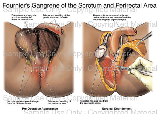
35-year-old male presented in the emergency department with the swelling over scrotum and watery discharge from the left side of the scrotum. Initially he developed a small swelling at the root of one of hair on the left side of the scrotum. Gradually the area around the swelling became red, painful and hard. The redness, swelling and the pain spread over whole of the scrotum within 2 days and the affected hair follicle started discharging watery fluid. Patient became feverish, dull and pale. On examination, there was gross involvement of the scrotum, which was swollen, inflamed and severely tender on touch (Figure1).

On examination there was gross thickening of the scrotal skin and testis were not palpable. Ultrasonography showed a scrotal wall thickening of 1 cm with collection of hyper echoic fluid around the testes. Doppler showed normal blood supply to both the testes.
Patient shifted to emergency operation theater and whole of the infected scrotal wall debrided exposing the testes. Daily dressing done with povidone iodine for 9 days after which the testes covered with the healthy granulation tissue (Figure 2).
On the 9th day in the elective surgery, after preparing the wound, excess granulation tissue scraped from the testes. The pedicle fascio cutaneous flaps developed from the femoral triangle on both the side as shown in the figure 3 by sharp dissection by using bipolar cautary.

The flaps rotated and tied over the bare testes in the midline and fixed with staples. Then the upper margins of the flaps fixed to the upper margin of the raw area finally followed by the lower margin. Flaps rotated and placed without tension with corrugated drains underneath. (Figure 4).

The area left behind on the femoral triangles on both the thighs due to the rotation of the flaps covered with split thickness skin graft taken from the lower anterior portion of the right thigh (Figure 5).

The drains were removed on the third postoperative day. The flaps were taken up well and the spit thickness skin graft uptake over the residual raw area were good. The patient sent home on tenth day and was under follow up lately until last 1 year without any complications.


