Causes, Symptoms and sings of aortic valvular insufficiency

A 74-year-old man presents with exertional dyspnea and generalized weakness. On examination, you discover a high-pitched, blowing diastolic murmur and a wide pulse pressure with bounding pulses. The most likely diagnosis is?
  • A) aortic stenosis
  • B) aortic insufficiency
  • C) mitral stenosis
  • D) mitral insufficiency
  • E) coarctation of the aorta

Answer and Discussion
The answer is B.
Causes of aortic regurgitation :
The prevalence of aortic regurgitation increases with age. Unlike aortic valve stenosis, aortic valvular insufficiency is rarely caused by degenerative aortic valve disease. Acute aortic valvular insufficiency may be due to infective endocarditis, aortic dissection, trauma, or rupture of the sinus of Valsalva. Chronic aortic insufficiency can be caused by valve leaflet disease, including rheumatic heart disease, congenital heart disease, rheumatoid arthritis, ankylosing spondylitis, or myxomatous degeneration. Chronic aortic insufficiency may also be caused by aortic root disease secondary to systemic hypertension, syphilitic aortitis, cystic medial necrosis, ankylosing spondylitis, rheumatoid arthritis, Reiter's disease, systemic lupus erythematosus, Ehlers–Danlos syndrome, and pseudoxanthoma elasticum.

Symptoms and sings of aortic valvular insufficiency :
The symptoms of aortic valvular insufficiency are the same in older persons as they are in younger ones. Usually, the main symptoms are related to heart failure, with exertional dyspnea and weakness being common symptoms. In some elderly patients, symptoms of dyspnea and palpitations may be more common at rest than with exertion. Nocturnal angina pectoris, often accompanied by flushing, diaphoresis, and palpitations, may occur; this is thought to be related to the slowing of the heart rate and the drop of arterial diastolic pressure. The classic findings of a high-pitched, blowing diastolic murmur and a wide pulse pressure with an abruptly rising and collapsing pulse should make the diagnosis of aortic valvular insufficiency easily recognized in elderly patients.

Photo description of Pterygium of the eye

Pterygium is a raised, wedge-shaped, non-cancerous growth of the conjunctiva onto the cornea, characterized by elastotic degeneration of and fibrovascular proliferation
Pterygium has a characteristic shape as it is triangular, with the base of the triangle located in the conjunctiva and the apex of the triangle encroaching onto the cornea.
With corneal involvement, even if arrested surgically, a pterygium can affect vision by warping the surface of the cornea and inducing astigmatism, and/or by actually growing over in front of the pupil and obstructing the entering light.
 

Triangular, thin, transparent conjunctival fold in the palpebral fissure. Its head is yellow, avascular and pints toward the corneal center. The body of the pterygium extends to the semilunar fold(this image for a 32-year-old man).

Treatment :
Pterygia seem to occur more frequently in people who spent much time outside, and is especially common in the southern lattitudes "Some consider pterygium as an occupational disease" So protecting the eyes from sun, dust and wind and use of lubricating drops is the standard treatment.
If the pterygium is still troublesome it can be removed surgically under local anaesthetic ,with high rate of recurrence. Once the pterygium is removed, the bare sclera is covered with conjunctiva

Photos and information about Blue Nevus

- Blue Nevus is a group of melanocytic lesions which all appear blue in colour due to the optical effects of light reflecting off melanin deep in the dermis.

- The blue nevus is a benign, usually solitary lesion, represents a localized proliferation of dermal melanocytes. It presents as a dark blue to black, moderately firm, rounded, sharply defined nodular tumour composed of spindle-shaped melanocytes with slender cytoplasmic processes, occurring often in association with melanin-laden macrophages in a sclerotic dermis.
- A blue naevus (nevus) is a rather unusual but non-cancerous mole.

- The blue nevus is also called the blue skin mole, or Jadassohn-Tièche nevus with 2 clinically recognized variants : the common blue nevus and the cellular blue nevus.
- Cellular blue nevus is larger, especially on buttocks and can degenerate into malignant melanoma.

- Clinically The blue nevus appears as a dark-blue or blue-black smooth nevus formed by melanin-heavily pigmented spindle cells in the middle and lower two-thirds of the dermis. Also, the blue nevus appears as a slate blue or bluish black, sharply circumscribed, flat or slightly elevated nodule, occurring on any area of the body.
- A biopsy should be performed on any changing pigmented lesion. For a solitary lesion, simple excision is usually curative. Rare cases of persistent blue nevi, manifesting as satellite lesions around the original excision site, have been reported. These must be distinguished from malignant blue nevus, and reexcision is recommended.

Common blue nevus

Atlas of Pathology images of Fibrocystic disease changes

A mass characterized by fibrocystic change is a pathological rather than a clinical diagnosis. A fibrocystic mass is diagnosed based on the presence of 2 important features: fibrous connective tissue and cysts

Fibrocystic changes are defined as Cystic changes and apocrine metaplasia involving terminal duct lobular units (TDLU) of breast tissue.

This image shows Extensive fibrocystic changes in serially sectioned formalin fixed breast tissue :
Note that the cysts are of various size are interspersed by dense fibrous tissue. This patient, who was in a high risk category for breast carcinoma, elected to have bilateral mastectomies with prosthetic implants. This degree of "fibrocystic change" becomes impossible to follow on mammograms due to complex shadows from the dense fibrous tissue.

Another gross picture for breast tissue with fibrocystic changes :


Note presence of "Blue dome" wich is a benign cysts filled by serous fluid often have this blue color when viewed from the outside


The next image is for Microscopic picture of Fibrocystic changes, including cystic dilatation, apocrine metaplasia, florid ductal hyperplasia, and fibrosis.


Grades of deep tendon reflexes

 Which of the following grades of deep tendon reflexes best describes a patient with transient
and intermittent clonus?
  • a.Grade 0
  • b.Grade 1+
  • c.Grade 2+
  • d.Grade 3+
  • e.Grade 4+

The answer is (e).
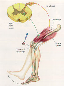
Clonus is rapidly alternating involuntary contraction and relaxation of skeletal muscle. Using a reflex hammer deep tendon reflex (DTR) response is graded on a scale from 0 to 4+:


In a normal person, when a muscle tendon is tapped briskly, the muscle immediately contracts due to a two-neuron reflex arc involving the spinal or brainstem segment that innervates the muscle. The afferent neuron whose cell body lies in a dorsal root ganglion innervates the muscle or Golgi tendon organ associated with the muscles; the efferent neuron is an alpha motoneuron in the anterior horn of the cord.

KSHV Infection & the Biology of Kaposi's Sarcoma

Donald E. Ganem, University of California, San Francisco and HHMI

Diastasis Recti in newborns


A vertical bulge down the midline of the abdomen can be seen in many newborns when intra-abdominal pressure increases. In this photo, a shadow lateral to the bulge can be seen going up the left side of the abdomen, starting near the umbilical clamp. Diastasis recti is caused by a relative weakness of linea alba "the fascia between the two rectus abdominus muscles".
It is essentially a cosmetic condition, with no associated morbidity or mortality .As it is not a herniation and is not pathologic, No treatment is needed ..... with time, this will disappear.
Another newborn has more pronounced diastasis recti. Spontaneous resolution is still expected. 

PVR or Proliferative Vitreo-Retinopathy (with video of Endoscopic Membranectomy)

In patients with Retinal Detachment "RD" ,Approximately 8-10% will develop Proliferative vitreoretinopathy (PVR) as a most common complication. Proliferative vitreoretinopathy is known simply as scar tissue formation that grows on the surface of the retina and pulls on the retina, causing it to redetach. The scar tissue also puckers the retina into stiff folds, like wrinkled aluminum foil.
Click on photo for enlargment

Unfold and reattach the retina is by removal the scar tissue with vitrectomy surgery and then reattach the retina. The lens of the eye almost always has to be removed during the surgery. If an intraocular lens implant is in the eye, it can be left alone.

PVR - Endoscopic Membranectomy


PVR or Proliferative Vitreo-Retinopathy (with video of Endoscopic Membranectomy)

B.R. of BrachioRadialis ;Medical mnemonic

BrachioRadialis:
Function: Its the Beer Raising muscle, flexes elbow, strongest when wrist is oriented like holding a beer.
Innervation: Breaks Rule: it's a flexor muscle, But Radial. (Radial nerve usually is for extensors: Recall BEST rule: B was for brachioradialis).
Important relation: Behind it is the Radial nerve in the cubital fossa.
Attachment: Attaches to Bottom of Radius.

Thoracic Kidney

A 74-year-old man with diabetes and hypertension was referred to the hospital for evaluation of a mass in the right side of the chest, identified on a radiograph (Panel A, arrows). He reported no pulmonary problems and no history of chest trauma. Physical examination was unremarkable. Reformatted coronal (Panel B) and sagittal (Panel C) computed tomographic scans show the right kidney (large arrows) and part of the liver protruding above the diaphragm (small arrows) and into the posteromedial aspect of the right hemithorax through the foramen of Bochdalek (arrowheads indicate the defective fusion of the diaphragm).

Thoracic kidney is a rare congenital anomaly. Because of the location of the liver, thoracic kidney on the right side is much less common than thoracic kidney on the left side. Thoracic kidney is twice as common in men as in women. Typically, the presence of a thoracic kidney is asymptomatic and requires no intervention, as in this case.

Triad of Kartagener syndrome

 A 14-year-old boy presents with a history of chronic sinusitis and frequent pneumonias. On physical examination, the patient has normal vital signs and is afebrile. He has mild frontal and maxillary sinus tenderness with palpation. Transillumination of the sinuses is normal. Heart sounds are best heard on the right side of the chest. The boy is coughing copious amounts of yellowish sputum. Which of the following is the most likely diagnosis?
  • a.Cystic fibrosis
  • b.Kartagener syndrome
  • c.Pulmonary dysplasia
  • d.Tuberculosis
  • e.Pulmonary hypertension

 The answer is b."Kartagener syndrome"
Kartagener syndrome is the inheritable disorder of dextrocardia, chronic sinusitis (with the formation of nasal polyps), and bronchiectasis. Patients may also present with situs inversus.The disorder is due to a defect that causes the cilia within the respiratory tract epithelium to become immotile. Cilia of the sperm are also affected.

Frontal and Chest x-ray showing dextrocardia "the typical radiographic sign of Kartagener's syndrom" and bronchiectasis.

Indication and Technique of Buried Intradermal Sutures

Indication:
Buried Intradermal Sutures technique is useful for wide, gaping wounds and when it is difficult to evert the skin edges. When buried intradermal sutures are placed properly, they make skin closure much easier. The purpose of this stitch is to line up the dermis and thus enhance healing.
It is termed buried as the knot needs to be as deep into the tissues as possible so that it does not come up through the epidermis and cause irritation and pain.

Technique:
-By using a cutting needle and absorbable material Start just under the dermal layer and come out below the epidermis.
You are going from deep to more superficial tissues.

-Now the technique becomes a bit challenging. You need to enter the skin on the opposite side at a depth similar to where you exited the skin on the first side, just below the epidermis. To do so, you should position the needle with the tip pointing down and pronate your wrist to get the correct angle. It will help to use the forceps (in the other hand) to hold up the skin. The needle should come out of the tissues below the dermis. Try to get as little fat in the stitch as possible; it does not contribute to the suture.

-Finnaly Tie the suture.


Indication and Technique of Buried Intradermal Sutures


Dumping syndrome after peptic ulcer surgery

A 44-year-old man with a history of peptic ulcer surgery presents with palpitations, tachycardia, lightheadedness, and diaphoresis after eating a meal. The symptoms typically begin 30 min after eating.Which of the following is the most likely diagnosis?
  • a.Malabsorption
  • b.Peptic ulcer recurrence
  • c.Gastric carcinoma
  • d.Gastritis
  • e.Dumping syndrome
  • f.Esophagitis

The answer is (e).


Some patients with a history of ulcer surgery experience the dumping syndrome 30 min after eating. They present with palpitations, tachycardia, lightheadedness, and diaphoresis after eating a meal due to the rapid emptying of hyperosmolar gastric contents into the small intestine.
Dumping syndrome is also called rapid gastric emptying which occurs when the undigested food bypass the  stomach and transported or "dumped" into the small intestine too rapidly.

Myringotomy tube insertion

Example of myringotomy and tube placement. Tubes are inserted for chronic are signifcant recurrent ear infections. They typically stay several months to a couple of years. They are intended to ventilate the middle ear space in order to treat as well as prevent future middle ear infections.

Atypical "Rodent Ulcer" Basal Cell Carcinoma

These lesions are moist ulcers which may not have the characteristic rolled, translucent border of a nodular Rodent Ulcer or Basal Cell Carcinoma BCC. Often, they are much larger than the pre-auricular lesion seen here, and represent neglected lesions.

The typical basal cell carcinoma appears as a small, pearly, dome-shaped nodule with small visible blood vessels (telangiectasias).